6OVT

Crystal Structure of IlvD from Mycobacterium tuberculosis


Experimental Data Snapshot

  • Method: X-RAY DIFFRACTION
  • Resolution: 1.88 Å
  • R-Value Free: 0.201 
  • R-Value Work: 0.160 
  • R-Value Observed: 0.162 

wwPDB Validation   3D Report Full Report


This is version 1.2 of the entry. See complete history


Literature

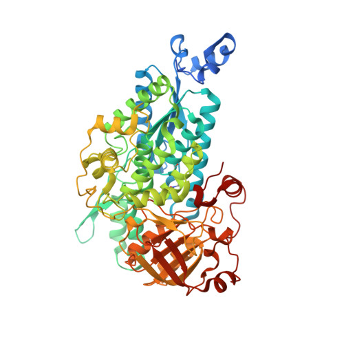
The active site of theMycobacterium tuberculosisbranched-chain amino acid biosynthesis enzyme dihydroxyacid dehydratase contains a 2Fe-2S cluster.

Bashiri, G.Grove, T.L.Hegde, S.S.Lagautriere, T.Gerfen, G.J.Almo, S.C.Squire, C.J.Blanchard, J.S.Baker, E.N.

(2019) J Biol Chem 294: 13158-13170

  • DOI: https://doi.org/10.1074/jbc.RA119.009498
  • Primary Citation of Related Structures:  
    6OVT

  • PubMed Abstract: 

    Iron-sulfur clusters are protein cofactors with an ancient evolutionary origin. These clusters are best known for their roles in redox proteins such as ferredoxins, but some iron-sulfur clusters have nonredox roles in the active sites of enzymes. Such clusters are often prone to oxidative degradation, making the enzymes difficult to characterize. Here we report a structural and functional characterization of dihydroxyacid dehydratase (DHAD) from Mycobacterium tuberculosis ( Mtb ), an essential enzyme in the biosynthesis of branched-chain amino acids. Conducting this analysis under fully anaerobic conditions, we solved the DHAD crystal structure, at 1.88 Å resolution, revealing a 2Fe-2S cluster in which one iron ligand is a potentially exchangeable water molecule or hydroxide. UV and EPR spectroscopy both suggested that the substrate binds directly to the cluster or very close to it. Kinetic analysis implicated two ionizable groups in the catalytic mechanism, which we postulate to be Ser-491 and the iron-bound water/hydroxide. Site-directed mutagenesis showed that Ser-491 is essential for activity, and substrate docking indicated that this residue is perfectly placed for proton abstraction. We found that a bound Mg 2+ ion 6.5 Å from the 2Fe-2S cluster plays a key role in substrate binding. We also identified a putative entry channel that enables access to the cluster and show that Mtb -DHAD is inhibited by a recently discovered herbicide, aspterric acid, that, given the essentiality of DHAD for Mtb survival, is a potential lead compound for the design of novel anti-TB drugs.


  • Organizational Affiliation

    Maurice Wilkins Center for Molecular Biodiscovery and School of Biological Sciences, University of Auckland, Auckland 1010, New Zealand.


Macromolecules
Find similar proteins by:  (by identity cutoff)  |  3D Structure
Entity ID: 1
MoleculeChains Sequence LengthOrganismDetailsImage
Dihydroxy-acid dehydratase
A, B, C, D
600Mycobacterium tuberculosisMutation(s): 0 
Gene Names: 
EC: 4.2.1.9
UniProt
Find proteins for P9WKJ5 (Mycobacterium tuberculosis (strain ATCC 25618 / H37Rv))
Explore P9WKJ5 
Go to UniProtKB:  P9WKJ5
Entity Groups  
Sequence Clusters30% Identity50% Identity70% Identity90% Identity95% Identity100% Identity
UniProt GroupP9WKJ5
Sequence Annotations
Expand
  • Reference Sequence
Small Molecules
Ligands 4 Unique
IDChains Name / Formula / InChI Key2D Diagram3D Interactions
FES
Query on FES

Download Ideal Coordinates CCD File 
F [auth A],
K [auth B],
O [auth C],
V [auth D]
FE2/S2 (INORGANIC) CLUSTER
Fe2 S2
NIXDOXVAJZFRNF-UHFFFAOYSA-N
PEG
Query on PEG

Download Ideal Coordinates CCD File 
I [auth A],
L [auth B],
P [auth C]
DI(HYDROXYETHYL)ETHER
C4 H10 O3
MTHSVFCYNBDYFN-UHFFFAOYSA-N
MG
Query on MG

Download Ideal Coordinates CCD File 
E [auth A]
G [auth A]
H [auth A]
J [auth B]
M [auth B]
E [auth A],
G [auth A],
H [auth A],
J [auth B],
M [auth B],
N [auth B],
Q [auth C],
R [auth C],
S [auth C],
T [auth D],
U [auth D],
W [auth D]
MAGNESIUM ION
Mg
JLVVSXFLKOJNIY-UHFFFAOYSA-N
NA
Query on NA

Download Ideal Coordinates CCD File 
X [auth D]SODIUM ION
Na
FKNQFGJONOIPTF-UHFFFAOYSA-N
Modified Residues  1 Unique
IDChains TypeFormula2D DiagramParent
KCX
Query on KCX
A, B, C, D
L-PEPTIDE LINKINGC7 H14 N2 O4LYS
Experimental Data & Validation

Experimental Data

  • Method: X-RAY DIFFRACTION
  • Resolution: 1.88 Å
  • R-Value Free: 0.201 
  • R-Value Work: 0.160 
  • R-Value Observed: 0.162 
  • Space Group: P 61
Unit Cell:
Length ( Å )Angle ( ˚ )
a = 88.406α = 90
b = 88.406β = 90
c = 483.307γ = 120
Software Package:
Software NamePurpose
REFMACrefinement
XDSdata reduction
Aimlessdata scaling
SHELXphasing
PDB_EXTRACTdata extraction

Structure Validation

View Full Validation Report


Entry History & Funding Information

Deposition Data


Funding OrganizationLocationGrant Number
National Institutes of Health/National Institute Of Allergy and Infectious Diseases (NIH/NIAID)United StatesAI133329
National Institutes of Health/National Institute of General Medical Sciences (NIH/NIGMS)United StatesU54-GM093342

Revision History  (Full details and data files)

  • Version 1.0: 2019-08-07
    Type: Initial release
  • Version 1.1: 2019-09-11
    Changes: Data collection, Database references
  • Version 1.2: 2019-12-18
    Changes: Author supporting evidence