6U6N

Structure of the trimeric globular domain of Adiponectin mutant - D187A Q188A


Experimental Data Snapshot

  • Method: X-RAY DIFFRACTION
  • Resolution: 2.15 Å
  • R-Value Free: 0.256 
  • R-Value Work: 0.210 
  • R-Value Observed: 0.214 

wwPDB Validation   3D Report Full Report


This is version 1.2 of the entry. See complete history


Literature

Mapping and engineering the interaction between adiponectin and T-cadherin.

Pascolutti, R.Erlandson, S.C.Burri, D.J.Zheng, S.Kruse, A.C.

(2020) J Biol Chem 295: 2749-2759

  • DOI: https://doi.org/10.1074/jbc.RA119.010970
  • Primary Citation of Related Structures:  
    6U66, 6U6N

  • PubMed Abstract: 

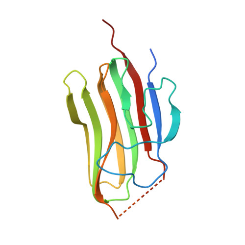
    Adiponectin is a highly abundant protein hormone secreted by adipose tissue. It elicits diverse biological responses, including anti-diabetic, anti-inflammatory, anti-tumor, and anti-atherosclerotic effects. Adiponectin consists of a globular domain and a collagen-like domain, and it occurs in three major oligomeric forms that self-assemble: trimers, hexamers, and high-molecular-weight oligomers. Adiponectin has been reported to bind to two seven-transmembrane domain receptors, AdipoR1 and AdipoR2, as well as to the protein T-cadherin, which is highly expressed in the cardiovascular system and binds only the high-molecular-weight form of adiponectin. The molecular mechanisms underlying this specificity remain unclear. Here we used a combination of X-ray crystallography and protein engineering to define the details of adiponectin's interaction with T-cadherin. We found that T-cadherin binds to the globular domain of adiponectin, relying on structural stabilization of this domain by bound metal ions. Moreover, we show that the adiponectin globular domain can be engineered to enhance its binding affinity for T-cadherin. These results help to define the molecular basis for the interaction between adiponectin and T-cadherin, and our engineered globular domain variants may be useful tools for further investigating adiponectin's functions.


  • Organizational Affiliation

    Department of Biological Chemistry and Molecular Pharmacology, Blavatnik Institute, Harvard Medical School, Boston, Massachusetts 02115.


Macromolecules
Find similar proteins by:  (by identity cutoff)  |  3D Structure
Entity ID: 1
MoleculeChains Sequence LengthOrganismDetailsImage
AdiponectinA [auth C]143Homo sapiensMutation(s): 2 
Gene Names: ADIPOQACDCACRP30APM1GBP28
UniProt & NIH Common Fund Data Resources
Find proteins for Q15848 (Homo sapiens)
Explore Q15848 
Go to UniProtKB:  Q15848
PHAROS:  Q15848
GTEx:  ENSG00000181092 
Entity Groups  
Sequence Clusters30% Identity50% Identity70% Identity90% Identity95% Identity100% Identity
UniProt GroupQ15848
Sequence Annotations
Expand
  • Reference Sequence
Small Molecules
Ligands 1 Unique
IDChains Name / Formula / InChI Key2D Diagram3D Interactions
CL
Query on CL

Download Ideal Coordinates CCD File 
B [auth C]CHLORIDE ION
Cl
VEXZGXHMUGYJMC-UHFFFAOYSA-M
Experimental Data & Validation

Experimental Data

  • Method: X-RAY DIFFRACTION
  • Resolution: 2.15 Å
  • R-Value Free: 0.256 
  • R-Value Work: 0.210 
  • R-Value Observed: 0.214 
  • Space Group: H 3
Unit Cell:
Length ( Å )Angle ( ˚ )
a = 75.704α = 90
b = 75.704β = 90
c = 48.865γ = 120
Software Package:
Software NamePurpose
PHENIXrefinement
PDB_EXTRACTdata extraction
XDSdata reduction
XSCALEdata scaling
PHASERphasing

Structure Validation

View Full Validation Report


Entry History & Funding Information

Deposition Data


Funding OrganizationLocationGrant Number
National Institutes of Health/National Human Genome Research Institute (NIH/NHGRI)United StatesDP5OD021345

Revision History  (Full details and data files)

  • Version 1.0: 2020-01-22
    Type: Initial release
  • Version 1.1: 2020-03-11
    Changes: Database references
  • Version 1.2: 2023-10-11
    Changes: Data collection, Database references, Refinement description