6R2W

Crystal structure of the super-active FVIIa variant VYT in complex with tissue factor


Experimental Data Snapshot

  • Method: X-RAY DIFFRACTION
  • Resolution: 1.25 Å
  • R-Value Free: 0.194 
  • R-Value Work: 0.170 
  • R-Value Observed: 0.171 

wwPDB Validation   3D Report Full Report


Ligand Structure Quality Assessment 


This is version 2.1 of the entry. See complete history


Literature

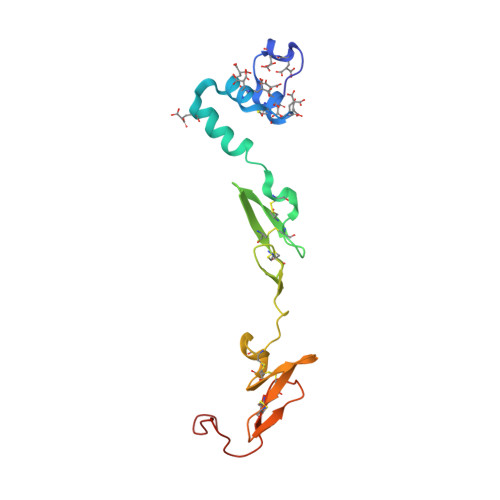
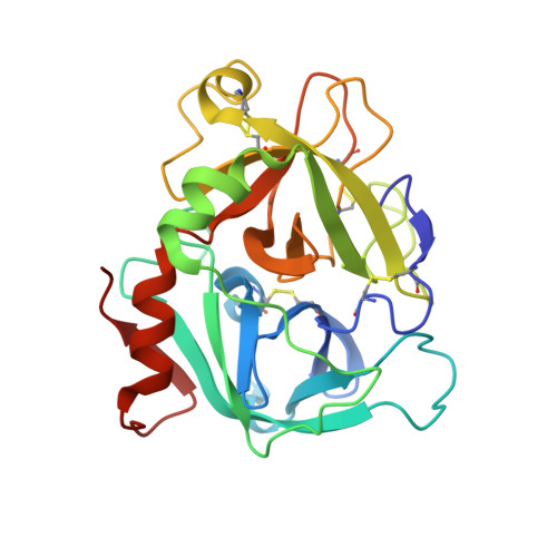
Beating tissue factor at its own game: Design and properties of a soluble tissue factor-independent coagulation factor VIIa.

Sorensen, A.B.Tuneew, I.Svensson, L.A.Persson, E.Ostergaard, H.Overgaard, M.T.Olsen, O.H.Gandhi, P.S.

(2020) J Biol Chem 295: 517-528

  • DOI: https://doi.org/10.1074/jbc.RA119.009183
  • Primary Citation of Related Structures:  
    6R2W

  • PubMed Abstract: 

    Two decades of research have uncovered the mechanism by which the complex of tissue factor (TF) and the plasma serine protease factor VIIa (FVIIa) mediates the initiation of blood coagulation. Membrane-anchored TF directly interacts with substrates and induces allosteric effects in the protease domain of FVIIa. These properties are also recapitulated by the soluble ectodomain of TF (sTF). At least two interdependent allosteric activation pathways originate at the FVIIa:sTF interface are proposed to enhance FVIIa activity upon sTF binding. Here, we sought to engineer an sTF-independent FVIIa variant by stabilizing both proposed pathways, with one pathway terminating at segment 215-217 in the activation domain and the other pathway terminating at the N terminus insertion site. To stabilize segment 215-217, we replaced the flexible 170 loop of FVIIa with the more rigid 170 loop from trypsin and combined it with an L163V substitution (FVIIa-VY T ). The FVIIa-VYT variant exhibited 60-fold higher amidolytic activity than FVIIa, and displayed similar FX activation and antithrombin inhibition kinetics to the FVIIa.sTF complex. The sTF-independent activity of FVIIa-VY T was partly mediated by an increase in the N terminus insertion and, as shown by X-ray crystallography, partly by Tyr-172 inserting into a cavity in the activation domain stabilizing the S1 substrate-binding pocket. The combination with L163V likely drove additional changes in a delicate hydrogen-bonding network that further stabilized S1-S3 sites. In summary, we report the first FVIIa variant that is catalytically independent of sTF and provide evidence supporting the existence of two TF-mediated allosteric activation pathways.


  • Organizational Affiliation

    Global Research, Novo Nordisk A/S, DK-2760 Måløv, Denmark; Department of Chemistry and Bioscience, Aalborg University, DK-9220 Aalborg, Denmark. Electronic address: abs@evaxion-biotech.com.


Macromolecules
Find similar proteins by:  (by identity cutoff)  |  3D Structure
Entity ID: 1
MoleculeChains Sequence LengthOrganismDetailsImage
Coagulation factor VIIA [auth L]143Homo sapiensMutation(s): 0 
Gene Names: F7
EC: 3.4.21.21
UniProt & NIH Common Fund Data Resources
Find proteins for P08709 (Homo sapiens)
Explore P08709 
Go to UniProtKB:  P08709
PHAROS:  P08709
GTEx:  ENSG00000057593 
Entity Groups  
Sequence Clusters30% Identity50% Identity70% Identity90% Identity95% Identity100% Identity
UniProt GroupP08709
Sequence Annotations
Expand
  • Reference Sequence
Find similar proteins by:  (by identity cutoff)  |  3D Structure
Entity ID: 2
MoleculeChains Sequence LengthOrganismDetailsImage
Coagulation factor VIIB [auth H]249Homo sapiensMutation(s): 7 
Gene Names: F7
EC: 3.4.21.21
UniProt & NIH Common Fund Data Resources
Find proteins for P08709 (Homo sapiens)
Explore P08709 
Go to UniProtKB:  P08709
PHAROS:  P08709
GTEx:  ENSG00000057593 
Entity Groups  
Sequence Clusters30% Identity50% Identity70% Identity90% Identity95% Identity100% Identity
UniProt GroupP08709
Sequence Annotations
Expand
  • Reference Sequence
Find similar proteins by:  (by identity cutoff)  |  3D Structure
Entity ID: 3
MoleculeChains Sequence LengthOrganismDetailsImage
Tissue factorC [auth T]210Homo sapiensMutation(s): 0 
Gene Names: F3
UniProt & NIH Common Fund Data Resources
Find proteins for P13726 (Homo sapiens)
Explore P13726 
Go to UniProtKB:  P13726
PHAROS:  P13726
GTEx:  ENSG00000117525 
Entity Groups  
Sequence Clusters30% Identity50% Identity70% Identity90% Identity95% Identity100% Identity
UniProt GroupP13726
Sequence Annotations
Expand
  • Reference Sequence
Oligosaccharides

Help

Entity ID: 4
MoleculeChains Length2D Diagram Glycosylation3D Interactions
alpha-D-xylopyranose-(1-3)-alpha-D-xylopyranose-(1-3)-beta-D-glucopyranoseD [auth A]3O-Glycosylation
Glycosylation Resources
GlyTouCan:  G41179DF
GlyCosmos:  G41179DF
GlyGen:  G41179DF
Small Molecules
Ligands 4 Unique
IDChains Name / Formula / InChI Key2D Diagram3D Interactions
0Z7
Query on 0Z7

Download Ideal Coordinates CCD File 
P [auth H]N-acetyl-D-phenylalanyl-N-[(2S,3S)-6-carbamimidamido-1-chloro-2-hydroxyhexan-3-yl]-L-phenylalaninamide
C27 H38 Cl N6 O4
SPENPXIFXGPVHG-UARRHKHWSA-O
FUC
Query on FUC

Download Ideal Coordinates CCD File 
M [auth L]alpha-L-fucopyranose
C6 H12 O5
SHZGCJCMOBCMKK-SXUWKVJYSA-N
TMA
Query on TMA

Download Ideal Coordinates CCD File 
N [auth L]
Q [auth H]
R [auth H]
S [auth H]
T [auth H]
N [auth L],
Q [auth H],
R [auth H],
S [auth H],
T [auth H],
U [auth H],
V [auth T],
W [auth T],
X [auth T],
Y [auth T]
TETRAMETHYLAMMONIUM ION
C4 H12 N
QEMXHQIAXOOASZ-UHFFFAOYSA-N
CA
Query on CA

Download Ideal Coordinates CCD File 
E [auth L]
F [auth L]
G [auth L]
H [auth L]
I [auth L]
E [auth L],
F [auth L],
G [auth L],
H [auth L],
I [auth L],
J [auth L],
K [auth L],
L,
O [auth H]
CALCIUM ION
Ca
BHPQYMZQTOCNFJ-UHFFFAOYSA-N
Modified Residues  1 Unique
IDChains TypeFormula2D DiagramParent
CGU
Query on CGU
A [auth L]L-PEPTIDE LINKINGC6 H9 N O6GLU
Experimental Data & Validation

Experimental Data

  • Method: X-RAY DIFFRACTION
  • Resolution: 1.25 Å
  • R-Value Free: 0.194 
  • R-Value Work: 0.170 
  • R-Value Observed: 0.171 
  • Space Group: P 21 21 21
Unit Cell:
Length ( Å )Angle ( ˚ )
a = 71.31α = 90
b = 80.04β = 90
c = 123.38γ = 90
Software Package:
Software NamePurpose
PHENIXrefinement
XDSdata reduction
XSCALEdata scaling
PHASERphasing
PDB_EXTRACTdata extraction

Structure Validation

View Full Validation Report


Ligand Structure Quality Assessment 


Entry History 

Deposition Data

Revision History  (Full details and data files)

  • Version 1.0: 2019-12-11
    Type: Initial release
  • Version 1.1: 2019-12-18
    Changes: Database references
  • Version 1.2: 2020-01-22
    Changes: Database references
  • Version 2.0: 2020-07-29
    Type: Remediation
    Reason: Carbohydrate remediation
    Changes: Advisory, Atomic model, Data collection, Derived calculations, Refinement description, Structure summary
  • Version 2.1: 2024-01-24
    Changes: Data collection, Database references, Derived calculations, Refinement description, Structure summary