6N5W

Crystal structure of the Ca2+/CaM complex with independent peptides of Kv7.4 (KCNQ4) A & B domains


Experimental Data Snapshot

  • Method: X-RAY DIFFRACTION
  • Resolution: 2.15 Å
  • R-Value Free: 0.274 
  • R-Value Work: 0.233 
  • R-Value Observed: 0.237 

wwPDB Validation   3D Report Full Report

Currently 6N5W does not have a validation slider image.


This is version 1.3 of the entry. See complete history


Literature

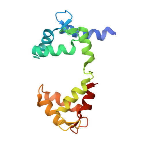
A mutually induced conformational fit underlies Ca2+-directed interactions between calmodulin and the proximal C terminus of KCNQ4 K+channels.

Archer, C.R.Enslow, B.T.Taylor, A.B.De la Rosa, V.Bhattacharya, A.Shapiro, M.S.

(2019) J Biol Chem 294: 6094-6112

  • DOI: https://doi.org/10.1074/jbc.RA118.006857
  • Primary Citation of Related Structures:  
    6N5W

  • PubMed Abstract: 

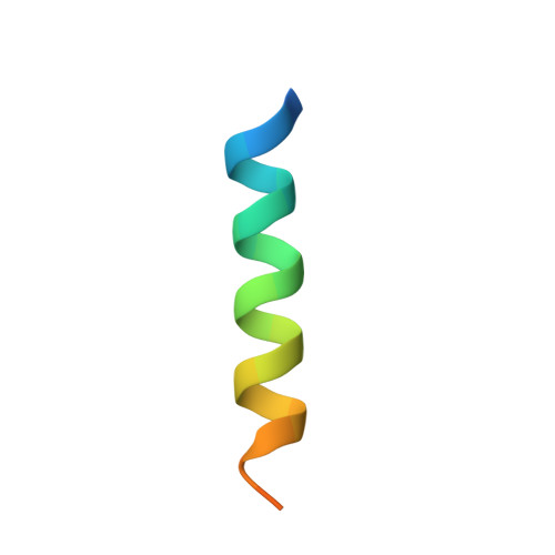
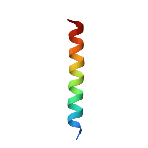
    Calmodulin (CaM) conveys intracellular Ca 2+ signals to KCNQ (Kv7, "M-type") K + channels and many other ion channels. Whether this "calmodulation" involves a dramatic structural rearrangement or only slight perturbations of the CaM/KCNQ complex is as yet unclear. A consensus structural model of conformational shifts occurring between low nanomolar and physiologically high intracellular [Ca 2+ ] is still under debate. Here, we used various techniques of biophysical chemical analyses to investigate the interactions between CaM and synthetic peptides corresponding to the A and B domains of the KCNQ4 subtype. We found that in the absence of CaM, the peptides are disordered, whereas Ca 2+ /CaM imposed helical structure on both KCNQ A and B domains. Isothermal titration calorimetry revealed that Ca 2+ /CaM has higher affinity for the B domain than for the A domain of KCNQ2-4 and much higher affinity for the B domain when prebound with the A domain. X-ray crystallography confirmed that these discrete peptides spontaneously form a complex with Ca 2+ /CaM, similar to previous reports of CaM binding KCNQ-AB domains that are linked together. Microscale thermophoresis and heteronuclear single-quantum coherence NMR spectroscopy indicated the C-lobe of Ca 2+ -free CaM to interact with the KCNQ4 B domain ( K d ∼10-20 μm), with increasing Ca 2+ molar ratios shifting the CaM-B domain interactions via only the CaM C-lobe to also include the N-lobe. Our findings suggest that in response to increased Ca 2+ , CaM undergoes lobe switching that imposes a dramatic mutually induced conformational fit to both the proximal C terminus of KCNQ4 channels and CaM, likely underlying Ca 2+ -dependent regulation of KCNQ gating.


  • Organizational Affiliation

    From the Departments of Cell and Integrative Physiology, University of Texas Health San Antonio, San Antonio, Texas 78229; Departments of Biochemistry and Structural Biology, University of Texas Health San Antonio, San Antonio, Texas 78229.


Macromolecules
Find similar proteins by:  (by identity cutoff)  |  3D Structure
Entity ID: 1
MoleculeChains Sequence LengthOrganismDetailsImage
Potassium voltage-gated channel subfamily KQT member 427Homo sapiensMutation(s): 0 
UniProt & NIH Common Fund Data Resources
Find proteins for P56696 (Homo sapiens)
Explore P56696 
Go to UniProtKB:  P56696
PHAROS:  P56696
GTEx:  ENSG00000117013 
Entity Groups  
Sequence Clusters30% Identity50% Identity70% Identity90% Identity95% Identity100% Identity
UniProt GroupP56696
Sequence Annotations
Expand
  • Reference Sequence
Find similar proteins by:  (by identity cutoff)  |  3D Structure
Entity ID: 2
MoleculeChains Sequence LengthOrganismDetailsImage
Potassium voltage-gated channel subfamily KQT member 426Homo sapiensMutation(s): 0 
UniProt & NIH Common Fund Data Resources
Find proteins for P56696 (Homo sapiens)
Explore P56696 
Go to UniProtKB:  P56696
PHAROS:  P56696
GTEx:  ENSG00000117013 
Entity Groups  
Sequence Clusters30% Identity50% Identity70% Identity90% Identity95% Identity100% Identity
UniProt GroupP56696
Sequence Annotations
Expand
  • Reference Sequence
Find similar proteins by:  (by identity cutoff)  |  3D Structure
Entity ID: 3
MoleculeChains Sequence LengthOrganismDetailsImage
Calmodulin-1149Homo sapiensMutation(s): 0 
Gene Names: CALM1CALMCAMCAM1
UniProt & NIH Common Fund Data Resources
Find proteins for P0DP23 (Homo sapiens)
Explore P0DP23 
Go to UniProtKB:  P0DP23
PHAROS:  P0DP23
GTEx:  ENSG00000198668 
Entity Groups  
Sequence Clusters30% Identity50% Identity70% Identity90% Identity95% Identity100% Identity
UniProt GroupP0DP23
Sequence Annotations
Expand
  • Reference Sequence
Experimental Data & Validation

Experimental Data

  • Method: X-RAY DIFFRACTION
  • Resolution: 2.15 Å
  • R-Value Free: 0.274 
  • R-Value Work: 0.233 
  • R-Value Observed: 0.237 
  • Space Group: P 21 21 2
Unit Cell:
Length ( Å )Angle ( ˚ )
a = 43.51α = 90
b = 130.57β = 90
c = 36.16γ = 90
Software Package:
Software NamePurpose
PHENIXrefinement
XDSdata reduction
XDSdata scaling
PHASERphasing

Structure Validation

View Full Validation Report

Currently 6N5W does not have a validation slider image.


Entry History & Funding Information

Deposition Data


Funding OrganizationLocationGrant Number
National Institutes of Health/National Institute of Neurological Disorders and Stroke (NIH/NINDS)United StatesR01 NS043394
National Institutes of Health/National Institute of Neurological Disorders and Stroke (NIH/NINDS)United StatesR01 NS094461
National Institutes of Health/National Heart, Lung, and Blood Institute (NIH/NHLBI)United StatesT32 HL007446

Revision History  (Full details and data files)

  • Version 1.0: 2019-03-13
    Type: Initial release
  • Version 1.1: 2019-04-24
    Changes: Data collection, Database references
  • Version 1.2: 2019-12-04
    Changes: Author supporting evidence
  • Version 1.3: 2023-10-11
    Changes: Data collection, Database references, Refinement description