6MAV

Complex of tissue inhibitor of metalloproteinase-1 (TIMP-1) mutant L34G with matrix metalloproteinase-3 catalytic domain (MMP-3cd)


Experimental Data Snapshot

  • Method: X-RAY DIFFRACTION
  • Resolution: 2.37 Å
  • R-Value Free: 0.298 
  • R-Value Work: 0.217 
  • R-Value Observed: 0.221 

wwPDB Validation   3D Report Full Report


This is version 1.3 of the entry. See complete history


Literature

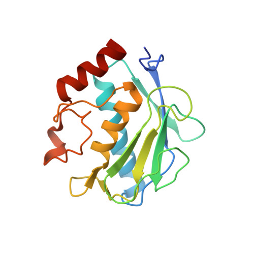
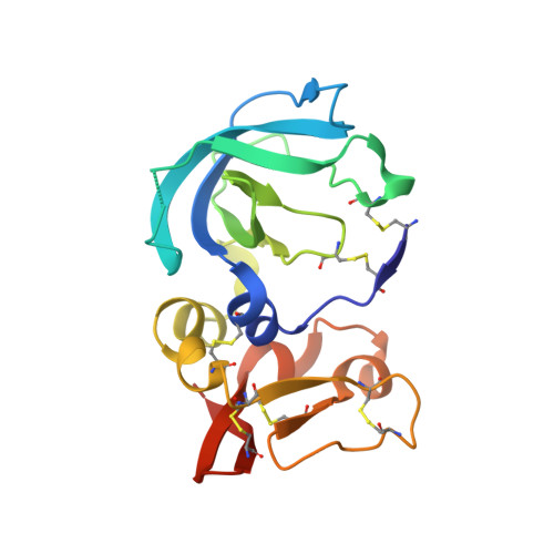
Directed evolution of the metalloproteinase inhibitor TIMP-1 reveals that its N- and C-terminal domains cooperate in matrix metalloproteinase recognition.

Raeeszadeh-Sarmazdeh, M.Greene, K.A.Sankaran, B.Downey, G.P.Radisky, D.C.Radisky, E.S.

(2019) J Biol Chem 294: 9476-9488

  • DOI: https://doi.org/10.1074/jbc.RA119.008321
  • Primary Citation of Related Structures:  
    6MAV, 6N9D

  • PubMed Abstract: 

    Tissue inhibitors of metalloproteinases (TIMPs) are natural inhibitors of matrix metalloproteinases (MMPs), enzymes that contribute to cancer and many inflammatory and degenerative diseases. The TIMP N-terminal domain binds and inhibits an MMP catalytic domain, but the role of the TIMP C-terminal domain in MMP inhibition is poorly understood. Here, we employed yeast surface display for directed evolution of full-length human TIMP-1 to develop MMP-3-targeting ultrabinders. By simultaneously incorporating diversity into both domains, we identified TIMP-1 variants that were up to 10-fold improved in binding MMP-3 compared with WT TIMP-1, with inhibition constants ( K i ) in the low picomolar range. Analysis of individual and paired mutations from the selected TIMP-1 variants revealed cooperative effects between distant residues located on the N- and C-terminal TIMP domains, positioned on opposite sides of the interaction interface with MMP-3. Crystal structures of MMP-3 complexes with TIMP-1 variants revealed conformational changes in TIMP-1 near the cooperative mutation sites. Affinity was strengthened by cinching of a reciprocal "tyrosine clasp" formed between the N-terminal domain of TIMP-1 and proximal MMP-3 interface and by changes in secondary structure within the TIMP-1 C-terminal domain that stabilize interdomain interactions and improve complementarity to MMP-3. Our protein engineering and structural studies provide critical insight into the cooperative function of TIMP domains and the significance of peripheral TIMP epitopes in MMP recognition. Our findings suggest new strategies to engineer TIMP proteins for therapeutic applications, and our directed evolution approach may also enable exploration of functional domain interactions in other protein systems.


  • Organizational Affiliation

    From the Department of Cancer Biology, Mayo Clinic, Jacksonville, Florida 32224.


Macromolecules
Find similar proteins by:  (by identity cutoff)  |  3D Structure
Entity ID: 1
MoleculeChains Sequence LengthOrganismDetailsImage
Stromelysin-1168Homo sapiensMutation(s): 0 
Gene Names: MMP3STMY1
EC: 3.4.24.17
UniProt & NIH Common Fund Data Resources
Find proteins for P08254 (Homo sapiens)
Explore P08254 
Go to UniProtKB:  P08254
PHAROS:  P08254
GTEx:  ENSG00000149968 
Entity Groups  
Sequence Clusters30% Identity50% Identity70% Identity90% Identity95% Identity100% Identity
UniProt GroupP08254
Sequence Annotations
Expand
  • Reference Sequence
Find similar proteins by:  (by identity cutoff)  |  3D Structure
Entity ID: 2
MoleculeChains Sequence LengthOrganismDetailsImage
Metalloproteinase inhibitor 1184Homo sapiensMutation(s): 1 
Gene Names: TIMP1CLGITIMP
UniProt & NIH Common Fund Data Resources
Find proteins for P01033 (Homo sapiens)
Explore P01033 
Go to UniProtKB:  P01033
PHAROS:  P01033
GTEx:  ENSG00000102265 
Entity Groups  
Sequence Clusters30% Identity50% Identity70% Identity90% Identity95% Identity100% Identity
UniProt GroupP01033
Sequence Annotations
Expand
  • Reference Sequence
Experimental Data & Validation

Experimental Data

  • Method: X-RAY DIFFRACTION
  • Resolution: 2.37 Å
  • R-Value Free: 0.298 
  • R-Value Work: 0.217 
  • R-Value Observed: 0.221 
  • Space Group: P 65 2 2
Unit Cell:
Length ( Å )Angle ( ˚ )
a = 69.703α = 90
b = 69.703β = 90
c = 321.326γ = 120
Software Package:
Software NamePurpose
PHENIXrefinement
PDB_EXTRACTdata extraction
xia2data reduction
Aimlessdata scaling
MOLREPphasing

Structure Validation

View Full Validation Report


Entry History & Funding Information

Deposition Data


Funding OrganizationLocationGrant Number
National Institutes of Health/National Cancer Institute (NIH/NCI)United StatesCA205471
Department of Defense (DOD, United States)United StatesDOD W81XWH-16-2-0030

Revision History  (Full details and data files)

  • Version 1.0: 2019-05-15
    Type: Initial release
  • Version 1.1: 2019-06-26
    Changes: Data collection, Database references
  • Version 1.2: 2019-11-27
    Changes: Author supporting evidence
  • Version 1.3: 2023-10-11
    Changes: Data collection, Database references, Derived calculations, Refinement description