6HPR

Crystal structure of cIAP1 RING domain bound to UbcH5B-Ub and a non-covalent Ub


Experimental Data Snapshot

  • Method: X-RAY DIFFRACTION
  • Resolution: 1.70 Å
  • R-Value Free: 0.197 
  • R-Value Work: 0.170 
  • R-Value Observed: 0.172 

wwPDB Validation   3D Report Full Report


This is version 1.6 of the entry. See complete history


Literature

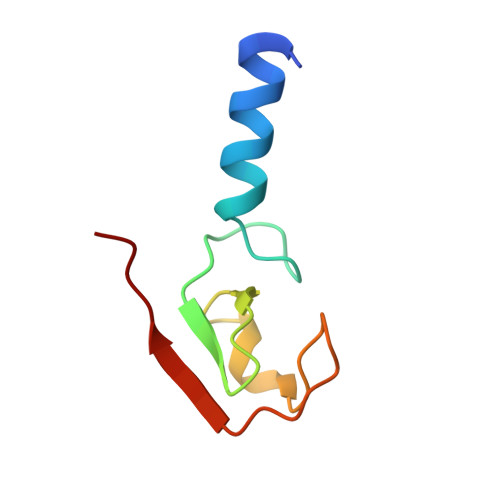
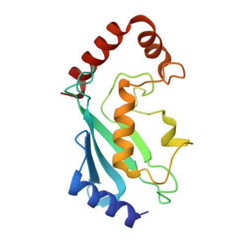
Structural insights into non-covalent ubiquitin activation of the cIAP1-UbcH5B∼ubiquitin complex.

Patel, A.Sibbet, G.J.Huang, D.T.

(2019) J Biol Chem 294: 1240-1249

  • DOI: https://doi.org/10.1074/jbc.RA118.006045
  • Primary Citation of Related Structures:  
    6HPR

  • PubMed Abstract: 

    Ubiquitin (Ub)-conjugating enzymes and Ub ligases control protein degradation and regulate many cellular processes in eukaryotes. Cellular inhibitor of apoptosis protein-1 (cIAP1) plays a central role in apoptosis and tumor necrosis factor signaling. It harbors a C-terminal RING domain that homodimerizes to recruit E2∼Ub (where ∼ denotes a thioester bond) complex to catalyze Ub transfer. Noncovalent Ub binding to the backside of the E2 Ub-conjugating enzyme UbcH5 has previously been shown to enhance RING domain activity, but the molecular basis for this enhancement is unclear. To investigate how dimeric cIAP1 RING activates E2∼Ub for Ub transfer and what role noncovalently bound Ub has in Ub transfer, here we determined the crystal structure of the cIAP1 RING dimer bound to both UbcH5B covalently linked to Ub (UbcH5B-Ub) and a noncovalent Ub to 1.7 Å resolution. The structure along with biochemical analyses revealed that the cIAP1 RING domain interacts with UbcH5B-Ub and thereby promotes the formation of a closed UbcH5B-Ub conformation that primes the thioester bond for Ub transfer. We observed that the noncovalent Ub binds to the backside of UbcH5B and abuts UbcH5B's α1β1-loop, which, in turn, stabilizes the closed UbcH5B-Ub conformation. Our results disclose the mechanism by which cIAP1 RING dimer activates UbcH5B∼Ub and indicate that noncovalent Ub binding further stabilizes the cIAP1-UbcH5B∼Ub complex in the active conformation to stimulate Ub transfer.


  • Organizational Affiliation

    Cancer Research UK Beatson Institute, Garscube Estate, Switchback Road, Glasgow G61 1BD, Scotland, United Kingdom; Institute of Cancer Sciences, University of Glasgow, Glasgow G61 1BD, Scotland, United Kingdom.


Macromolecules
Find similar proteins by:  (by identity cutoff)  |  3D Structure
Entity ID: 1
MoleculeChains Sequence LengthOrganismDetailsImage
Baculoviral IAP repeat-containing protein 265Homo sapiensMutation(s): 0 
Gene Names: BIRC2API1MIHBRNF48
EC: 2.3.2.27
UniProt & NIH Common Fund Data Resources
Find proteins for Q13490 (Homo sapiens)
Explore Q13490 
Go to UniProtKB:  Q13490
PHAROS:  Q13490
GTEx:  ENSG00000110330 
Entity Groups  
Sequence Clusters30% Identity50% Identity70% Identity90% Identity95% Identity100% Identity
UniProt GroupQ13490
Sequence Annotations
Expand
  • Reference Sequence
Find similar proteins by:  (by identity cutoff)  |  3D Structure
Entity ID: 2
MoleculeChains Sequence LengthOrganismDetailsImage
Polyubiquitin-B
B, D
81Homo sapiensMutation(s): 0 
Gene Names: UBB
UniProt & NIH Common Fund Data Resources
Find proteins for P0CG47 (Homo sapiens)
Explore P0CG47 
Go to UniProtKB:  P0CG47
PHAROS:  P0CG47
GTEx:  ENSG00000170315 
Entity Groups  
Sequence Clusters30% Identity50% Identity70% Identity90% Identity95% Identity100% Identity
UniProt GroupP0CG47
Sequence Annotations
Expand
  • Reference Sequence
Find similar proteins by:  (by identity cutoff)  |  3D Structure
Entity ID: 3
MoleculeChains Sequence LengthOrganismDetailsImage
Ubiquitin-conjugating enzyme E2 D2147Homo sapiensMutation(s): 0 
Gene Names: UBE2D2PUBC1UBC4UBC5BUBCH4UBCH5B
EC: 2.3.2.23 (PDB Primary Data), 2.3.2.24 (PDB Primary Data)
UniProt & NIH Common Fund Data Resources
Find proteins for P62837 (Homo sapiens)
Explore P62837 
Go to UniProtKB:  P62837
PHAROS:  P62837
GTEx:  ENSG00000131508 
Entity Groups  
Sequence Clusters30% Identity50% Identity70% Identity90% Identity95% Identity100% Identity
UniProt GroupP62837
Sequence Annotations
Expand
  • Reference Sequence
Experimental Data & Validation

Experimental Data

  • Method: X-RAY DIFFRACTION
  • Resolution: 1.70 Å
  • R-Value Free: 0.197 
  • R-Value Work: 0.170 
  • R-Value Observed: 0.172 
  • Space Group: C 1 2 1
Unit Cell:
Length ( Å )Angle ( ˚ )
a = 79.19α = 90
b = 53.6β = 107.57
c = 78.54γ = 90
Software Package:
Software NamePurpose
PHENIXrefinement
xia2data reduction
xia2data scaling
PHASERphasing

Structure Validation

View Full Validation Report


Entry History & Funding Information

Deposition Data


Funding OrganizationLocationGrant Number
Cancer Research UKUnited KingdomA23278

Revision History  (Full details and data files)

  • Version 1.0: 2018-12-12
    Type: Initial release
  • Version 1.1: 2018-12-19
    Changes: Data collection, Database references
  • Version 1.2: 2018-12-26
    Changes: Data collection, Database references
  • Version 1.3: 2019-03-06
    Changes: Data collection, Database references
  • Version 1.4: 2019-05-08
    Changes: Advisory, Data collection, Derived calculations
  • Version 1.5: 2019-07-10
    Changes: Data collection
  • Version 1.6: 2024-01-24
    Changes: Data collection, Database references, Refinement description