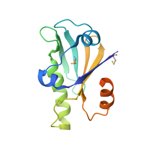
The crystal structure of theHelicobacter pyloriLlaJI.R1 N-terminal domain provides a model for site-specific DNA binding.
Hosford, C.J., Chappie, J.S.(2018) J Biol Chem 293: 11758-11771
- PubMed: 29895618
- DOI: https://doi.org/10.1074/jbc.RA118.001888
- Primary Citation of Related Structures:
6C5D - PubMed Abstract:
Restriction modification systems consist of an endonuclease that cleaves foreign DNA site-specifically and an associated methyltransferase that protects the corresponding target site in the host genome. Modification-dependent restriction systems, in contrast, specifically recognize and cleave methylated and/or glucosylated DNA. The LlaJI restriction system contains two 5-methylcytosine (5mC) methyltransferases (LlaJI.M1 and LlaJI.M2) and two restriction proteins (LlaJI.R1 and LlaJI.R2). LlaJI.R1 and LlaJI.R2 are homologs of McrB and McrC, respectively, which in Escherichia coli function together as a modification-dependent restriction complex specific for 5mC-containing DNA. Lactococcus lactis LlaJI.R1 binds DNA site-specifically, suggesting that the LlaJI system uses a different mode of substrate recognition. Here we present the structure of the N-terminal DNA-binding domain of Helicobacter pylori LlaJI.R1 at 1.97-Å resolution, which adopts a B3 domain fold. Structural comparison to B3 domains in plant transcription factors and other restriction enzymes identifies key recognition motifs responsible for site-specific DNA binding. Moreover, biochemistry and structural modeling provide a rationale for how H. pylori LlaJI.R1 may bind a target site that differs from the 5-bp sequence recognized by other LlaJI homologs and identify residues critical for this recognition activity. These findings underscore the inherent structural plasticity of B3 domains, allowing recognition of a variety of substrates using the same structural core.
Organizational Affiliation:
From the Department of Molecular Medicine, Cornell University, Ithaca, New York 14853.