5G4A

AadA in complex with ATP and magnesium


Experimental Data Snapshot

  • Method: X-RAY DIFFRACTION
  • Resolution: 1.90 Å
  • R-Value Free: 0.222 
  • R-Value Work: 0.176 
  • R-Value Observed: 0.178 

wwPDB Validation   3D Report Full Report


Ligand Structure Quality Assessment 


This is version 2.1 of the entry. See complete history


Literature

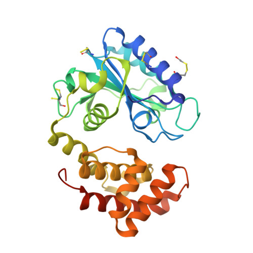
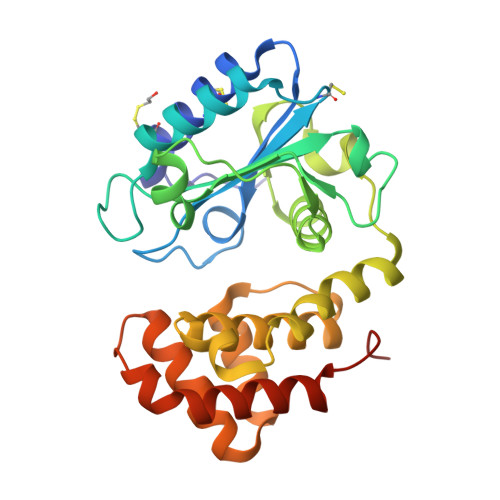
Structural mechanism of AadA, a dual-specificity aminoglycoside adenylyltransferase fromSalmonella enterica.

Stern, A.L.Van der Verren, S.E.Kanchugal P, S.Nasvall, J.Gutierrez-de-Teran, H.Selmer, M.

(2018) J Biol Chem 293: 11481-11490

  • DOI: https://doi.org/10.1074/jbc.RA118.003989
  • Primary Citation of Related Structures:  
    5G4A, 5LPA, 5LUH, 6FZB

  • PubMed Abstract: 

    Streptomycin and spectinomycin are antibiotics that bind to the bacterial ribosome and perturb protein synthesis. The clinically most prevalent bacterial resistance mechanism is their chemical modification by aminoglycoside-modifying enzymes such as aminoglycoside nucleotidyltransferases (ANTs). AadA from Salmonella enterica is an aminoglycoside (3″)(9) adenylyltransferase that O- adenylates position 3″ of streptomycin and position 9 of spectinomycin. We previously reported the apo-AadA structure with a closed active site. To clarify how AadA binds ATP and its two chemically distinct drug substrates, we here report crystal structures of WT AadA complexed with ATP, magnesium, and streptomycin and of an active-site mutant, E87Q, complexed with ATP and streptomycin or the closely related dihydrostreptomycin. These structures revealed that ATP binding induces a conformational change that positions the two domains for drug binding at the interdomain cleft and disclosed the interactions between both domains and the three rings of streptomycin. Spectinomycin docking followed by molecular dynamics simulations suggested that, despite the limited structural similarities with streptomycin, spectinomycin makes similar interactions around the modification site and, in agreement with mutational data, forms critical interactions with fewer residues. Using structure-guided sequence analyses of ANT(3″)(9) enzymes acting on both substrates and ANT(9) enzymes active only on spectinomycin, we identified sequence determinants for activity on each substrate. We experimentally confirmed that Trp-173 and Asp-178 are essential only for streptomycin resistance. Activity assays indicated that Glu-87 is the catalytic base in AadA and that the nonadenylating E87Q mutant can hydrolyze ATP in the presence of streptomycin.


  • Organizational Affiliation

    Department of Cell and Molecular Biology, Uppsala University, BMC, Box 596, SE-751 24 Uppsala, Sweden.


Macromolecules
Find similar proteins by:  (by identity cutoff)  |  3D Structure
Entity ID: 1
MoleculeChains Sequence LengthOrganismDetailsImage
Aminoglycoside (3'') (9) adenylyltransferase270Salmonella entericaMutation(s): 0 
Gene Names: aadA
UniProt
Find proteins for Q8ZPX9 (Salmonella typhimurium (strain LT2 / SGSC1412 / ATCC 700720))
Explore Q8ZPX9 
Go to UniProtKB:  Q8ZPX9
Entity Groups  
Sequence Clusters30% Identity50% Identity70% Identity90% Identity95% Identity100% Identity
UniProt GroupQ8ZPX9
Sequence Annotations
Expand
  • Reference Sequence
Find similar proteins by:  (by identity cutoff)  |  3D Structure
Entity ID: 2
MoleculeChains Sequence LengthOrganismDetailsImage
Aminoglycoside (3'') (9) adenylyltransferase270Salmonella entericaMutation(s): 0 
Gene Names: aadA
UniProt
Find proteins for Q8ZPX9 (Salmonella typhimurium (strain LT2 / SGSC1412 / ATCC 700720))
Explore Q8ZPX9 
Go to UniProtKB:  Q8ZPX9
Entity Groups  
Sequence Clusters30% Identity50% Identity70% Identity90% Identity95% Identity100% Identity
UniProt GroupQ8ZPX9
Sequence Annotations
Expand
  • Reference Sequence
Small Molecules
Ligands 7 Unique
IDChains Name / Formula / InChI Key2D Diagram3D Interactions
ATP
Query on ATP

Download Ideal Coordinates CCD File 
J [auth A],
V [auth B]
ADENOSINE-5'-TRIPHOSPHATE
C10 H16 N5 O13 P3
ZKHQWZAMYRWXGA-KQYNXXCUSA-N
PGE
Query on PGE

Download Ideal Coordinates CCD File 
N [auth B]TRIETHYLENE GLYCOL
C6 H14 O4
ZIBGPFATKBEMQZ-UHFFFAOYSA-N
PEG
Query on PEG

Download Ideal Coordinates CCD File 
C [auth A],
O [auth B]
DI(HYDROXYETHYL)ETHER
C4 H10 O3
MTHSVFCYNBDYFN-UHFFFAOYSA-N
EDO
Query on EDO

Download Ideal Coordinates CCD File 
D [auth A],
E [auth A],
F [auth A],
P [auth B],
Q [auth B]
1,2-ETHANEDIOL
C2 H6 O2
LYCAIKOWRPUZTN-UHFFFAOYSA-N
CL
Query on CL

Download Ideal Coordinates CCD File 
G [auth A],
R [auth B]
CHLORIDE ION
Cl
VEXZGXHMUGYJMC-UHFFFAOYSA-M
MG
Query on MG

Download Ideal Coordinates CCD File 
H [auth A],
I [auth A],
S [auth B],
T [auth B],
U [auth B]
MAGNESIUM ION
Mg
JLVVSXFLKOJNIY-UHFFFAOYSA-N
UNX
Query on UNX

Download Ideal Coordinates CCD File 
K [auth A]
L [auth A]
M [auth A]
W [auth B]
X [auth B]
K [auth A],
L [auth A],
M [auth A],
W [auth B],
X [auth B],
Y [auth B]
UNKNOWN ATOM OR ION
X
Modified Residues  2 Unique
IDChains TypeFormula2D DiagramParent
CME
Query on CME
A
L-PEPTIDE LINKINGC5 H11 N O3 S2CYS
CSS
Query on CSS
A
L-PEPTIDE LINKINGC3 H7 N O2 S2CYS
Experimental Data & Validation

Experimental Data

  • Method: X-RAY DIFFRACTION
  • Resolution: 1.90 Å
  • R-Value Free: 0.222 
  • R-Value Work: 0.176 
  • R-Value Observed: 0.178 
  • Space Group: P 32
Unit Cell:
Length ( Å )Angle ( ˚ )
a = 82.33α = 90
b = 82.33β = 90
c = 79.11γ = 120
Software Package:
Software NamePurpose
PHENIXrefinement
XDSdata reduction
XSCALEdata scaling
PHASERphasing

Structure Validation

View Full Validation Report


Ligand Structure Quality Assessment 


Entry History 

Deposition Data

Revision History  (Full details and data files)

  • Version 1.0: 2017-11-29
    Type: Initial release
  • Version 1.1: 2019-06-12
    Changes: Data collection, Database references, Derived calculations
  • Version 2.0: 2022-12-07
    Changes: Advisory, Atomic model, Data collection, Database references, Derived calculations, Non-polymer description, Source and taxonomy, Structure summary
  • Version 2.1: 2024-01-31
    Changes: Data collection, Refinement description