5T1I

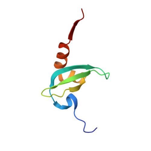
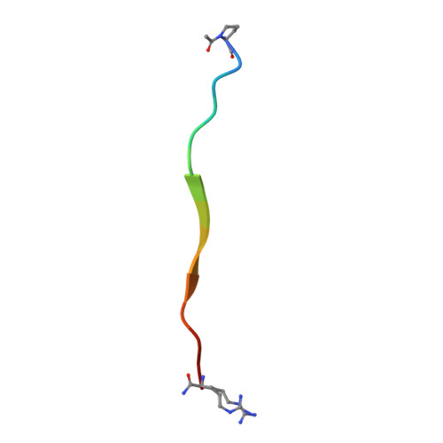
CBX3 chromo shadow domain in complex with histone H3 peptide


Experimental Data Snapshot

  • Method: X-RAY DIFFRACTION
  • Resolution: 1.60 Å
  • R-Value Free: 0.239 
  • R-Value Work: 0.194 
  • R-Value Observed: 0.197 

wwPDB Validation   3D Report Full Report


This is version 1.5 of the entry. See complete history


Literature

Peptide recognition by heterochromatin protein 1 (HP1) chromoshadow domains revisited: Plasticity in the pseudosymmetric histone binding site of human HP1.

Liu, Y.Qin, S.Lei, M.Tempel, W.Zhang, Y.Loppnau, P.Li, Y.Min, J.

(2017) J Biol Chem 292: 5655-5664

  • DOI: https://doi.org/10.1074/jbc.M116.768374
  • Primary Citation of Related Structures:  
    5T1I

  • PubMed Abstract: 

    Heterochromatin protein 1 (HP1), a highly conserved non-histone chromosomal protein in eukaryotes, plays important roles in the regulation of gene transcription. Each of the three human homologs of HP1 includes a chromoshadow domain (CSD). The CSD interacts with various proteins bearing the P X V X L motif but also with a region of histone H3 that bears the similar P XX V X L motif. The latter interaction has not yet been resolved in atomic detail. Here we demonstrate that the CSDs of all three human HP1 homologs have comparable affinities to the P XX V X L motif of histone H3. The HP1 C-terminal extension enhances the affinity, as does the increasing length of the H3 peptide. The crystal structure of the human HP1γ CSD (CSD γ ) in complex with an H3 peptide suggests that recognition of H3 by CSD γ to some extent resembles CSD-P X V X L interaction. Nevertheless, the prolyl residue of the P XX V X L motif appears to play a role distinct from that of Pro in the known HP1β CSD-P X V X L complexes. We consequently generalize the historical CSD-P X V X L interaction model and expand the search scope for additional CSD binding partners.


  • Organizational Affiliation

    From the Structural Genomics Consortium, University of Toronto, Toronto, Ontario M5G 1L7, Canada and.


Macromolecules
Find similar proteins by:  (by identity cutoff)  |  3D Structure
Entity ID: 1
MoleculeChains Sequence LengthOrganismDetailsImage
Chromobox protein homolog 3
A, B
68Homo sapiensMutation(s): 0 
Gene Names: CBX3
UniProt & NIH Common Fund Data Resources
Find proteins for Q13185 (Homo sapiens)
Explore Q13185 
Go to UniProtKB:  Q13185
PHAROS:  Q13185
GTEx:  ENSG00000122565 
Entity Groups  
Sequence Clusters30% Identity50% Identity70% Identity90% Identity95% Identity100% Identity
UniProt GroupQ13185
Sequence Annotations
Expand
  • Reference Sequence

Find similar proteins by:  Sequence   |   3D Structure  

Entity ID: 2
MoleculeChains Sequence LengthOrganismDetailsImage
Histone H3.115Homo sapiensMutation(s): 0 
UniProt & NIH Common Fund Data Resources
Find proteins for P68431 (Homo sapiens)
Explore P68431 
Go to UniProtKB:  P68431
PHAROS:  P68431
Entity Groups  
Sequence Clusters30% Identity50% Identity70% Identity90% Identity95% Identity100% Identity
UniProt GroupP68431
Sequence Annotations
Expand
  • Reference Sequence
Small Molecules
Ligands 1 Unique
IDChains Name / Formula / InChI Key2D Diagram3D Interactions
UNX
Query on UNX

Download Ideal Coordinates CCD File 
D [auth A]
E [auth A]
F [auth A]
G [auth B]
H [auth B]
D [auth A],
E [auth A],
F [auth A],
G [auth B],
H [auth B],
I [auth B],
J [auth B],
K [auth B],
L [auth B],
M [auth B],
N [auth C],
O [auth C]
UNKNOWN ATOM OR ION
X
Modified Residues  2 Unique
IDChains TypeFormula2D DiagramParent
AAR
Query on AAR
C
L-PEPTIDE LINKINGC6 H16 N5 OARG
N7P
Query on N7P
C
L-PEPTIDE LINKINGC7 H11 N O3PRO
Experimental Data & Validation

Experimental Data

Unit Cell:
Length ( Å )Angle ( ˚ )
a = 40.737α = 90
b = 34.971β = 93.32
c = 45.833γ = 90
Software Package:
Software NamePurpose
Aimlessdata scaling
REFMACrefinement
PDB_EXTRACTdata extraction
XDSdata scaling
PHASERphasing

Structure Validation

View Full Validation Report


Entry History 

Deposition Data

Revision History  (Full details and data files)

  • Version 1.0: 2016-09-14
    Type: Initial release
  • Version 1.1: 2016-10-12
    Changes: Structure summary
  • Version 1.2: 2016-10-19
    Changes: Database references
  • Version 1.3: 2017-03-08
    Changes: Database references
  • Version 1.4: 2017-04-19
    Changes: Database references
  • Version 1.5: 2023-10-04
    Changes: Data collection, Database references, Refinement description