5JY7

Complex of Mycobacterium smegmatis trehalose synthase with maltokinase


Experimental Data Snapshot

  • Method: X-RAY DIFFRACTION
  • Resolution: 3.60 Å
  • R-Value Free: 0.281 
  • R-Value Work: 0.261 
  • R-Value Observed: 0.262 

wwPDB Validation   3D Report Full Report


This is version 1.3 of the entry. See complete history


Literature

Crystal structure of the TreS-Pep2 complex, initiating alpha-glucan synthesis in the GlgE pathway of mycobacteria.

Kermani, A.A.Roy, R.Gopalasingam, C.Kocurek, K.I.Patel, T.R.Alderwick, L.J.Besra, G.S.Futterer, K.

(2019) J Biol Chem 

  • DOI: https://doi.org/10.1074/jbc.RA118.004297
  • Primary Citation of Related Structures:  
    5JY7

  • PubMed Abstract: 

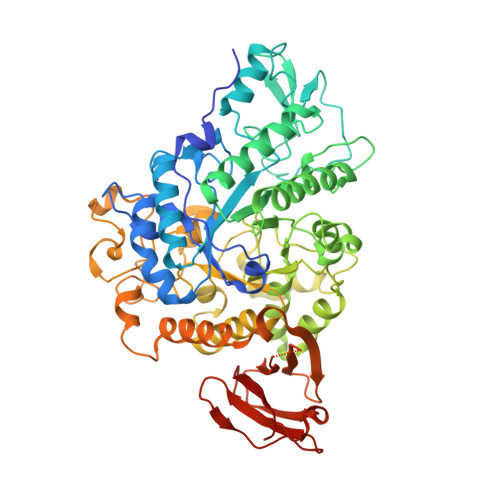
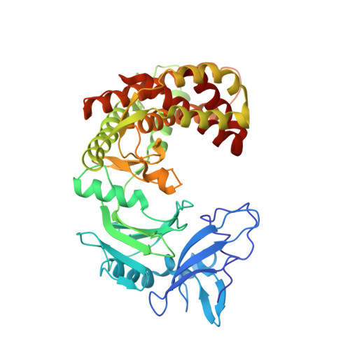
    A growing body of evidence implicates the mycobacterial capsule, the outermost layer of the mycobacterial cell envelope, in modulation of the host immune response and virulence of mycobacteria. Mycobacteria synthesize the dominant capsule component, α(1→4)-linked glucan, via three interconnected and potentially redundant metabolic pathways. Here, we report the crystal structure of the Mycobacterium smegmatis TreS:Pep2 complex, containing trehalose synthase (TreS) and maltokinase (Pep2), which converts trehalose to maltose 1-phosphate as part of the TreS:Pep2-GlgE pathway. The structure, at 3.6 Å resolution, revealed that a diamond-shaped TreS tetramer forms the core of the complex and that pairs of Pep2 monomers bind to opposite apices of the tetramer in a 4 + 4 configuration. However, for the M. smegmatis orthologues, results from isothermal titration calorimetry and analytical ultracentrifugation experiments indicated that the prevalent stoichiometry in solution is 4 TreS + 2 Pep2 protomers. The observed discrepancy between the crystallized complex and the behavior in the solution state may be explained by the relatively weak affinity of Pep2 for TreS ( K d 3.5 μm at mildly acidic pH) and crystal packing favoring the 4 + 4 complex. Proximity of the ATP-binding site in Pep2 to the complex interface provides a rational basis for rate enhancement of Pep2 upon binding to TreS, but the complex structure appears to rule out substrate channeling between the active sites of TreS and Pep2. Our findings provide a structural model for the trehalose synthase:maltokinase complex in M. smegmatis that offers critical insights into capsule assembly.


  • Organizational Affiliation

    From the Institute of Microbiology & Infection, School of Biosciences, University of Birmingham, Edgbaston, Birmingham B15 2TT, United Kingdom.


Macromolecules
Find similar proteins by:  (by identity cutoff)  |  3D Structure
Entity ID: 1
MoleculeChains Sequence LengthOrganismDetailsImage
Trehalose synthase/amylase TreS
A, B, C, D, E
A, B, C, D, E, F, G, H
593Mycolicibacterium smegmatis MC2 155Mutation(s): 0 
Gene Names: treSMSMEG_6515MSMEI_6343
EC: 3.2.1.1 (PDB Primary Data), 5.4.99.16 (PDB Primary Data)
UniProt
Find proteins for A0R6E0 (Mycolicibacterium smegmatis (strain ATCC 700084 / mc(2)155))
Explore A0R6E0 
Go to UniProtKB:  A0R6E0
Entity Groups  
Sequence Clusters30% Identity50% Identity70% Identity90% Identity95% Identity100% Identity
UniProt GroupA0R6E0
Sequence Annotations
Expand
  • Reference Sequence
Find similar proteins by:  (by identity cutoff)  |  3D Structure
Entity ID: 2
MoleculeChains Sequence LengthOrganismDetailsImage
Maltokinase
I, J, K, L, M
I, J, K, L, M, N, O, P
441Mycolicibacterium smegmatis MC2 155Mutation(s): 0 
Gene Names: makpep2MSMEG_6514MSMEI_6342
EC: 2.7.1.175
UniProt
Find proteins for A0R6D9 (Mycolicibacterium smegmatis (strain ATCC 700084 / mc(2)155))
Explore A0R6D9 
Go to UniProtKB:  A0R6D9
Entity Groups  
Sequence Clusters30% Identity50% Identity70% Identity90% Identity95% Identity100% Identity
UniProt GroupA0R6D9
Sequence Annotations
Expand
  • Reference Sequence
Experimental Data & Validation

Experimental Data

  • Method: X-RAY DIFFRACTION
  • Resolution: 3.60 Å
  • R-Value Free: 0.281 
  • R-Value Work: 0.261 
  • R-Value Observed: 0.262 
  • Space Group: P 43
Unit Cell:
Length ( Å )Angle ( ˚ )
a = 315.68α = 90
b = 315.68β = 90
c = 124.95γ = 90
Software Package:
Software NamePurpose
REFMACrefinement
PDB_EXTRACTdata extraction
XDSdata reduction
XSCALEdata scaling
PHASERphasing

Structure Validation

View Full Validation Report


Entry History & Funding Information

Deposition Data


Funding OrganizationLocationGrant Number
Medical Research Council (United Kingdom)United KingdomMR/K012118/1

Revision History  (Full details and data files)

  • Version 1.0: 2017-05-24
    Type: Initial release
  • Version 1.1: 2017-09-13
    Changes: Author supporting evidence
  • Version 1.2: 2019-04-24
    Changes: Data collection, Database references
  • Version 1.3: 2024-01-10
    Changes: Data collection, Database references, Refinement description