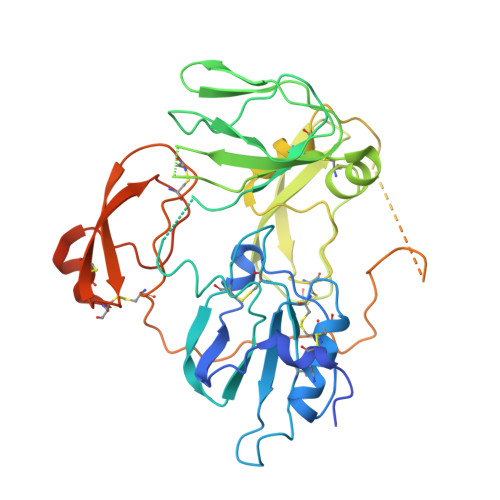
The crystal structure of a multidomain protease inhibitor (HAI-1) reveals the mechanism of its auto-inhibition
Liu, M., Yuan, C., Jensen, J.K., Zhao, B., Jiang, Y., Jiang, L., Huang, M.(2017) J Biol Chem 292: 8412-8423
- PubMed: 28348076
- DOI: https://doi.org/10.1074/jbc.M117.779256
- Primary Citation of Related Structures:
5H7V - PubMed Abstract:
Hepatocyte growth factor activator inhibitor 1 (HAI-1) is a membrane-bound multidomain protein essential to the integrity of the basement membrane during placental development and is also important in maintaining postnatal homeostasis in many tissues. HAI-1 is a Kunitz-type serine protease inhibitor, and soluble fragments of HAI-1 with variable lengths have been identified in vivo The full-length extracellular portion of HAI-1 (sHAI-1) shows weaker inhibitory activity toward target proteases than the smaller fragments, suggesting auto-inhibition of HAI-1. However, this possible regulatory mechanism has not yet been evaluated. Here, we solved the crystal structure of sHAI-1 and determined the solution structure by small-angle X-ray scattering. These structural analyses revealed that, despite the presence of long linkers, sHAI-1 exists in a compact conformation in which sHAI-1 active sites in Kunitz domain 1 are sterically blocked by neighboring structural elements. We also found that in the presence of target proteases, sHAI-1 adopts an extended conformation that disables the auto-inhibition effect. Our results also reveal the roles of non-inhibitory domains of this multidomain protein and explain the low activity of the full-length protein. The structural insights gained here improve our understanding of the regulation of HAI-1 inhibitory activities and point to new approaches for better controlling these activities.
Organizational Affiliation:
State Key Laboratory of Structural Chemistry, Fujian Institute of Research on the Structure of Matter, Chinese Academy of Sciences, Fuzhou, Fujian, 350002, China; University of Chinese Academy of Sciences, Beijing 100049, China.