5EAQ

Two active site divalent ion in the crystal structure of the hammerhead ribozyme bound to a transition state analog-Mn2+


Experimental Data Snapshot

  • Method: X-RAY DIFFRACTION
  • Resolution: 3.20 Å
  • R-Value Free: 0.278 
  • R-Value Work: 0.247 
  • R-Value Observed: 0.250 

wwPDB Validation   3D Report Full Report


This is version 1.1 of the entry. See complete history


Literature

Two Active Site Divalent Ions in the Crystal Structure of the Hammerhead Ribozyme Bound to a Transition State Analogue.

Mir, A.Golden, B.L.

(2016) Biochemistry 55: 633-636

  • DOI: https://doi.org/10.1021/acs.biochem.5b01139
  • Primary Citation of Related Structures:  
    5EAO, 5EAQ

  • PubMed Abstract: 

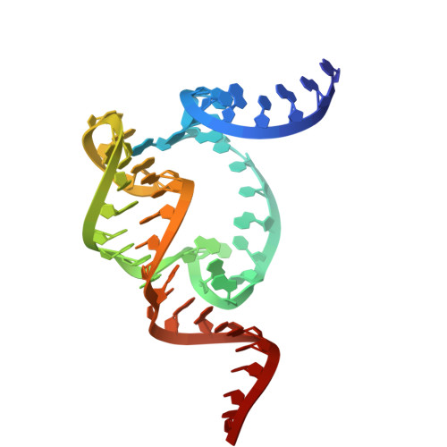
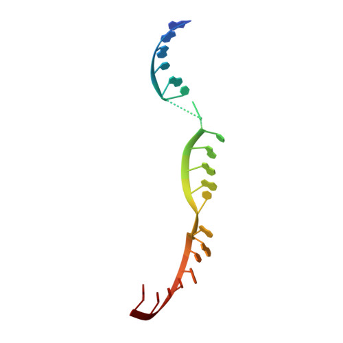
    The crystal structure of the hammerhead ribozyme bound to the pentavalent transition state analogue vanadate reveals significant rearrangements relative to the previously determined structures. The active site contracts, bringing G10.1 closer to the cleavage site and repositioning a divalent metal ion such that it could, ultimately, interact directly with the scissile phosphate. This ion could also position a water molecule to serve as a general acid in the cleavage reaction. A second divalent ion is observed coordinated to O6 of G12. This metal ion is well-placed to help tune the pKA of G12. On the basis of this crystal structure as well as a wealth of biochemical studies, we propose a mechanism in which G12 serves as the general base and a magnesium-bound water serves as a general acid.


  • Organizational Affiliation

    Department of Biochemistry, Purdue University , West Lafayette, Indiana 47907, United States.


Macromolecules
Find similar nucleic acids by:  (by identity cutoff)  |  3D Structure
Entity ID: 1
MoleculeChains LengthOrganismImage
RNA (48-MER)48synthetic construct
Sequence Annotations
Expand
  • Reference Sequence

Find similar nucleic acids by:  Sequence   |   3D Structure  

Entity ID: 2
MoleculeChains LengthOrganismImage
RNA (5'-R(*GP*GP*GP*CP*GP*U*(CVC)*UP*GP*GP*GP*CP*AP*GP*UP*AP*CP*CP*CP*A)-3')20synthetic construct
Sequence Annotations
Expand
  • Reference Sequence
Experimental Data & Validation

Experimental Data

  • Method: X-RAY DIFFRACTION
  • Resolution: 3.20 Å
  • R-Value Free: 0.278 
  • R-Value Work: 0.247 
  • R-Value Observed: 0.250 
  • Space Group: C 2 2 21
Unit Cell:
Length ( Å )Angle ( ˚ )
a = 81.251α = 90
b = 87.419β = 90
c = 105.539γ = 90
Software Package:
Software NamePurpose
PHENIXrefinement
HKL-2000data reduction
HKL-2000data scaling
PHENIXphasing

Structure Validation

View Full Validation Report


Entry History 

Deposition Data

Revision History  (Full details and data files)

  • Version 1.0: 2016-02-24
    Type: Initial release
  • Version 1.1: 2023-09-27
    Changes: Advisory, Data collection, Database references, Derived calculations, Refinement description