3CB4

The Crystal Structure of LepA


Experimental Data Snapshot

  • Method: X-RAY DIFFRACTION
  • Resolution: 2.80 Å
  • R-Value Free: 0.295 
  • R-Value Work: 0.246 
  • R-Value Observed: 0.249 

wwPDB Validation   3D Report Full Report


This is version 1.1 of the entry. See complete history


Literature

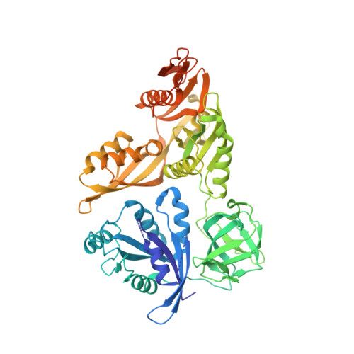
The structure of LepA, the ribosomal back translocase.

Evans, R.N.Blaha, G.Bailey, S.Steitz, T.A.

(2008) Proc Natl Acad Sci U S A 105: 4673-4678

  • DOI: https://doi.org/10.1073/pnas.0801308105
  • Primary Citation of Related Structures:  
    3CB4

  • PubMed Abstract: 

    LepA is a highly conserved elongation factor that promotes the back translocation of tRNAs on the ribosome during the elongation cycle. We have determined the crystal structure of LepA from Escherichia coli at 2.8-A resolution. The high degree of sequence identity between LepA and EF-G is reflected in the structural similarity between the individual homologous domains of LepA and EF-G. However, the orientation of domains III and V in LepA differs from their orientations in EF-G. LepA also contains a C-terminal domain (CTD) not found in EF-G that has a previously unobserved protein fold. The high structural similarity between LepA and EF-G enabled us to derive a homology model for LepA bound to the ribosome using a 7.3-A cryo-EM structure of a complex between EF-G and the 70S ribosome. In this model, the very electrostatically positive CTD of LepA is placed in the direct vicinity of the A site of the large ribosomal subunit, suggesting a possible interaction between the CTD and the back translocated tRNA or 23S rRNA.


  • Organizational Affiliation

    Departments of Molecular Biophysics and Biochemistry and Chemistry and Howard Hughes Medical Institute, Yale University, New Haven, CT 06520-8114, USA.


Macromolecules
Find similar proteins by:  (by identity cutoff)  |  3D Structure
Entity ID: 1
MoleculeChains Sequence LengthOrganismDetailsImage
GTP-binding protein lepA599Escherichia coli K-12Mutation(s): 0 
Gene Names: lepA
UniProt
Find proteins for P60785 (Escherichia coli (strain K12))
Explore P60785 
Go to UniProtKB:  P60785
Entity Groups  
Sequence Clusters30% Identity50% Identity70% Identity90% Identity95% Identity100% Identity
UniProt GroupP60785
Sequence Annotations
Expand
  • Reference Sequence
Experimental Data & Validation

Experimental Data

  • Method: X-RAY DIFFRACTION
  • Resolution: 2.80 Å
  • R-Value Free: 0.295 
  • R-Value Work: 0.246 
  • R-Value Observed: 0.249 
  • Space Group: P 1 21 1
Unit Cell:
Length ( Å )Angle ( ˚ )
a = 97.96α = 90
b = 146.243β = 100.6
c = 139.317γ = 90
Software Package:
Software NamePurpose
REFMACrefinement
CBASSdata collection
HKL-2000data reduction
SCALEPACKdata scaling
SOLVEphasing

Structure Validation

View Full Validation Report


Entry History 

Deposition Data

Revision History  (Full details and data files)

  • Version 1.0: 2008-03-18
    Type: Initial release
  • Version 1.1: 2011-07-13
    Changes: Advisory, Source and taxonomy, Version format compliance