3B39

Structure of the DnaG primase catalytic domain bound to ssDNA


Experimental Data Snapshot

  • Method: X-RAY DIFFRACTION
  • Resolution: 2.35 Å
  • R-Value Free: 0.254 
  • R-Value Work: 0.212 
  • R-Value Observed: 0.214 

wwPDB Validation   3D Report Full Report


This is version 1.3 of the entry. See complete history


Literature

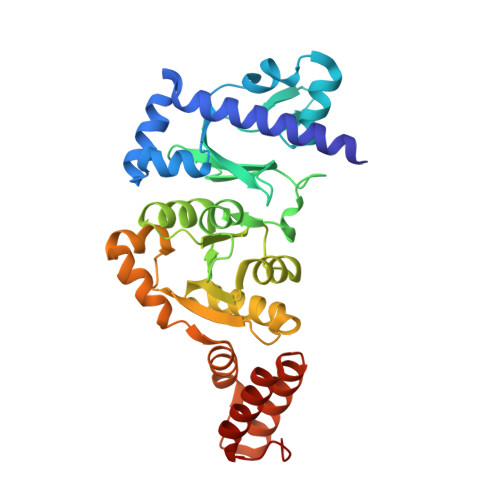
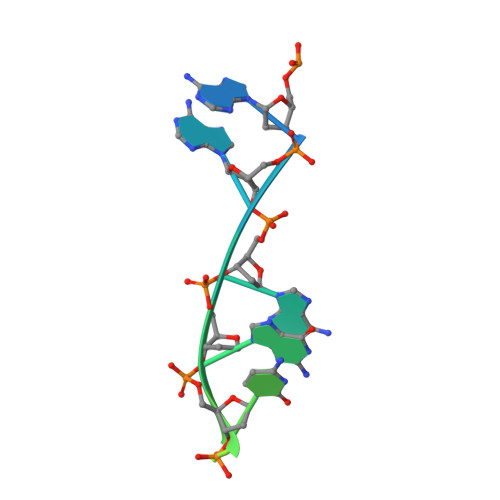
Identification of a DNA primase template tracking site redefines the geometry of primer synthesis.

Corn, J.E.Pelton, J.G.Berger, J.M.

(2008) Nat Struct Mol Biol 15: 163-169

  • DOI: https://doi.org/10.1038/nsmb.1373
  • Primary Citation of Related Structures:  
    3B39

  • PubMed Abstract: 

    Primases are essential RNA polymerases required for the initiation of DNA replication, lagging strand synthesis and replication restart. Many aspects of primase function remain unclear, including how the enzyme associates with a moving nucleic acid strand emanating from a helicase and orients primers for handoff to replisomal components. Using a new screening method to trap transient macromolecular interactions, we determined the structure of the Escherichia coli DnaG primase catalytic domain bound to single-stranded DNA. The structure reveals an unanticipated binding site that engages nucleic acid in two distinct configurations, indicating that it serves as a nonspecific capture and tracking locus for template DNA. Bioinformatic and biochemical analyses show that this evolutionarily constrained region enforces template polarity near the active site and is required for primase function. Together, our findings reverse previous proposals for primer-template orientation and reconcile disparate studies to re-evaluate replication fork organization.


  • Organizational Affiliation

    Department of Molecular and Cell Biology, QB3 Institute, 374D Stanley Hall no. 3220, University of California, Berkeley, Berkeley, California 94720, USA.


Macromolecules

Find similar proteins by:  (by identity cutoff)  |  3D Structure
Entity ID: 2
MoleculeChains Sequence LengthOrganismDetailsImage
DNA primaseC [auth A],
D [auth B]
322Escherichia coli K-12Mutation(s): 1 
Gene Names: dnaGdnaPparB
EC: 2.7.7
UniProt
Find proteins for P0ABS5 (Escherichia coli (strain K12))
Explore P0ABS5 
Go to UniProtKB:  P0ABS5
Entity Groups  
Sequence Clusters30% Identity50% Identity70% Identity90% Identity95% Identity100% Identity
UniProt GroupP0ABS5
Sequence Annotations
Expand
  • Reference Sequence

Find similar nucleic acids by:  Sequence   |   3D Structure  

Entity ID: 1
MoleculeChains LengthOrganismImage
DNA (5'-D(*DCP*DAP*DAP*DAP*DGP*DCP*DCP*DAP*DAP*DAP*DAP*DGP*DGP*DAP*DC)-3')A [auth C],
B [auth D]
15N/A
Sequence Annotations
Expand
  • Reference Sequence
Experimental Data & Validation

Experimental Data

  • Method: X-RAY DIFFRACTION
  • Resolution: 2.35 Å
  • R-Value Free: 0.254 
  • R-Value Work: 0.212 
  • R-Value Observed: 0.214 
  • Space Group: P 62
Unit Cell:
Length ( Å )Angle ( ˚ )
a = 136.829α = 90
b = 136.829β = 90
c = 71.071γ = 120
Software Package:
Software NamePurpose
REFMACrefinement
Blu-Icedata collection
HKL-2000data reduction
HKL-2000data scaling
PHASERphasing

Structure Validation

View Full Validation Report


Entry History 

Deposition Data

Revision History  (Full details and data files)

  • Version 1.0: 2008-01-15
    Type: Initial release
  • Version 1.1: 2011-07-13
    Changes: Advisory, Source and taxonomy, Version format compliance
  • Version 1.2: 2021-10-20
    Changes: Database references
  • Version 1.3: 2023-08-30
    Changes: Data collection, Refinement description