Expansion of Substrate Specificity and Catalytic Mechanism of Azoreductase by X-ray Crystallography and Site-directed Mutagenesis
Ito, K., Nakanishi, M., Lee, W.C., Zhi, Y., Sasaki, H., Zenno, S., Saigo, K., Kitade, Y., Tanokura, M.(2008) J Biol Chem 283: 13889-13896
- PubMed: 18337254
- DOI: https://doi.org/10.1074/jbc.M710070200
- Primary Citation of Related Structures:
2Z98, 2Z9B, 2Z9C, 2Z9D - PubMed Abstract:
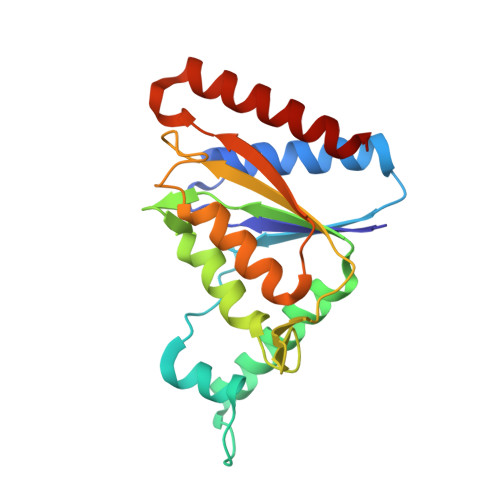
AzoR is an FMN-dependent NADH-azoreductase isolated from Escherichia coli as a protein responsible for the degradation of azo compounds. We previously reported the crystal structure of the enzyme in the oxidized form. In the present study, different structures of AzoR were determined under several conditions to obtain clues to the reaction mechanism of the enzyme. AzoR in its reduced form revealed a twisted butterfly bend of the isoalloxazine ring of the FMN cofactor and a rearrangement of solvent molecules. The crystal structure of oxidized AzoR in a different space group and the structure of the enzyme in complex with the inhibitor dicoumarol were also determined. These structures indicate that the formation of a hydrophobic part around the isoalloxazine ring is important for substrate binding and an electrostatic interaction between Arg-59 and the carboxyl group of the azo compound causes a substrate preference for methyl red over p-methyl red. The substitution of Arg-59 with Ala enhanced the Vmax value for p-methyl red 27-fold with a 3.8-fold increase of the Km value. This result indicates that Arg-59 decides the substrate specificity of AzoR. The Vmax value for the p-methyl red reduction of the R59A mutant is comparable with that for the methyl red reduction of the wild-type enzyme, whereas the activity toward methyl red was retained. These findings indicate the expansion of AzoR substrate specificity by a single amino acid substitution. Furthermore, we built an authentic model of the AzoR-methyl red complex based on the results of the study.
Organizational Affiliation:
Department of Applied Biological Chemistry, Graduate School of Agricultural and Life Sciences, The University of Tokyo, 1-1-1 Yayoi, Tokyo 113-8657, Japan.