


Crystal structure of periplasmic glycerophosphodiester phosphodiesterase from Escherichia coli
Malashkevich, V.M., Fedorov, E., Almo, S.C.To be published.
Experimental Data Snapshot
wwPDB Validation 3D Report Full Report

Entity ID: 1 | |||||
|---|---|---|---|---|---|
| Molecule | Chains | Sequence Length | Organism | Details | Image |
| Glycerophosphoryl diester phosphodiesterase | 356 | Escherichia coli | Mutation(s): 0 Gene Names: glpQ EC: 3.1.4.46 | 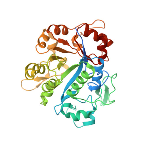 | |
UniProt | |||||
Find proteins for P09394 (Escherichia coli (strain K12)) Explore P09394 Go to UniProtKB: P09394 | |||||
Entity Groups | |||||
| Sequence Clusters | 30% Identity50% Identity70% Identity90% Identity95% Identity100% Identity | ||||
| UniProt Group | P09394 | ||||
Sequence AnnotationsExpand | |||||
| |||||
| Ligands 2 Unique | |||||
|---|---|---|---|---|---|
| ID | Chains | Name / Formula / InChI Key | 2D Diagram | 3D Interactions | |
| GOL Query on GOL | D [auth A], F [auth B] | GLYCEROL C3 H8 O3 PEDCQBHIVMGVHV-UHFFFAOYSA-N |  | ||
| CA Query on CA | C [auth A], E [auth B] | CALCIUM ION Ca BHPQYMZQTOCNFJ-UHFFFAOYSA-N |  | ||
| Length ( Å ) | Angle ( ˚ ) |
|---|---|
| a = 63.789 | α = 90 |
| b = 74.469 | β = 90 |
| c = 157.219 | γ = 90 |
| Software Name | Purpose |
|---|---|
| CNS | refinement |
| SCALEPACK | data scaling |
| AMoRE | phasing |
RCSB PDB (citation) is hosted by
RCSB PDB is a member of the

