1TRE

THE STRUCTURE OF TRIOSEPHOSPHATE ISOMERASE FROM ESCHERICHIA COLI DETERMINED AT 2.6 ANGSTROM RESOLUTION


Experimental Data Snapshot

  • Method: X-RAY DIFFRACTION
  • Resolution: 2.60 Å
  • R-Value Work: 0.119 
  • R-Value Observed: 0.119 

wwPDB Validation   3D Report Full Report


This is version 1.5 of the entry. See complete history


Literature

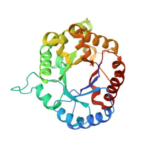
Structure of triosephosphate isomerase from Escherichia coli determined at 2.6 A resolution.

Noble, M.E.Zeelen, J.P.Wierenga, R.K.Mainfroid, V.Goraj, K.Gohimont, A.C.Martial, J.A.

(1993) Acta Crystallogr D Biol Crystallogr 49: 403-417

  • DOI: https://doi.org/10.1107/S0907444993002628
  • Primary Citation of Related Structures:  
    1TRE

  • PubMed Abstract: 

    The structure of triosephosphate isomerase (TIM) from the organism Escherichia coli has been determined at a resolution of 2.6 A. The structure was solved by the molecular replacement method, first at 2.8 A resolution with a crystal grown by the technique of hanging-drop crystallization from a mother liquor containing the transition-state analogue 2-phosphoglycolate (2PG). As a search model in the molecular replacement calculations, the refined structure of TIM from Trypanosoma brucei, which has a sequence identity of 46% compared to the enzyme from E. coli, was used. An E. coli TIM crystal grown in the absence of 2PG, diffracting to 2.6 A resolution, was later obtained by application of the technique of macro-seeding using a seed crystal grown from a mother liquor without 2PG. The final 2.6 A model has a crystallographic R factor of 11.9%, and agrees well with standard stereochemical parameters. The structure of E. coli TIM suggests the importance of residues which favour helix initiation for the formation of the TIM fold. In addition, TIM from E. coli shows peculiarities in its dimer interface, and in the packing of core residues within the beta-barrel.


  • Organizational Affiliation

    European Molecular Biology Laboratory, Heidelburg, Germany.


Macromolecules
Find similar proteins by:  (by identity cutoff)  |  3D Structure
Entity ID: 1
MoleculeChains Sequence LengthOrganismDetailsImage
TRIOSEPHOSPHATE ISOMERASE
A, B
255Escherichia coliMutation(s): 0 
EC: 5.3.1.1
UniProt
Find proteins for P0A858 (Escherichia coli (strain K12))
Explore P0A858 
Go to UniProtKB:  P0A858
Entity Groups  
Sequence Clusters30% Identity50% Identity70% Identity90% Identity95% Identity100% Identity
UniProt GroupP0A858
Sequence Annotations
Expand
  • Reference Sequence
Experimental Data & Validation

Experimental Data

  • Method: X-RAY DIFFRACTION
  • Resolution: 2.60 Å
  • R-Value Work: 0.119 
  • R-Value Observed: 0.119 
  • Space Group: P 21 21 21
Unit Cell:
Length ( Å )Angle ( ˚ )
a = 67.69α = 90
b = 46.78β = 90
c = 151.31γ = 90
Software Package:
Software NamePurpose
X-PLORmodel building
TNTrefinement
X-PLORrefinement
X-PLORphasing

Structure Validation

View Full Validation Report


Entry History 

Deposition Data

Revision History  (Full details and data files)

  • Version 1.0: 1993-10-31
    Type: Initial release
  • Version 1.1: 2008-03-24
    Changes: Version format compliance
  • Version 1.2: 2011-07-13
    Changes: Version format compliance
  • Version 1.3: 2019-07-17
    Changes: Data collection, Other, Refinement description
  • Version 1.4: 2019-08-14
    Changes: Data collection, Refinement description
  • Version 1.5: 2024-02-14
    Changes: Data collection, Database references