1QFJ

CRYSTAL STRUCTURE OF NAD(P)H:FLAVIN OXIDOREDUCTASE FROM ESCHERICHIA COLI


Experimental Data Snapshot

  • Method: X-RAY DIFFRACTION
  • Resolution: 2.20 Å
  • R-Value Free: 0.290 
  • R-Value Work: 0.239 
  • R-Value Observed: 0.239 

wwPDB Validation   3D Report Full Report


This is version 1.3 of the entry. See complete history


Literature

Crystal structure of NAD(P)H:flavin oxidoreductase from Escherichia coli.

Ingelman, M.Ramaswamy, S.Niviere, V.Fontecave, M.Eklund, H.

(1999) Biochemistry 38: 7040-7049

  • DOI: https://doi.org/10.1021/bi982849m
  • Primary Citation of Related Structures:  
    1QFJ

  • PubMed Abstract: 

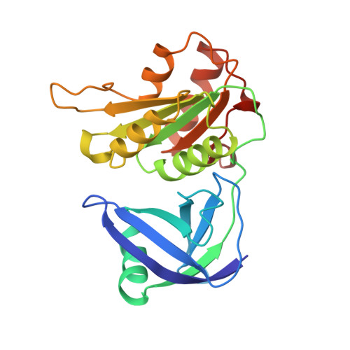
    Flavin reductases use flavins as substrates and are distinct from flavoenzymes which have tightly bound flavins. The reduced flavin can serve to reduce ferric complexes and iron proteins. In Escherichia coli, reactivation of ribonucleotide reductase is achieved by reduced flavins produced by flavin reductase. The crystal structure of E. coli flavin reductase reveals that the enzyme structure is similar to the structures of the ferredoxin reductase family of flavoproteins despite very low sequence similarities. The main difference between flavin reductase and structurally related flavoproteins is that there is no binding site for the AMP moiety of FAD. The direction of the helix in the flavin binding domain, corresponding to the phosphate binding helix in the flavoproteins, is also slightly different and less suitable for phosphate binding. Interactions for flavin substrates are instead provided by a hydrophobic isoalloxazine binding site that also contains a serine and a threonine, which form hydrogen bonds to the isoalloxazine of bound riboflavin in a substrate complex.


  • Organizational Affiliation

    Department of Molecular Biology, Swedish University of Agricultural Sciences, Biomedical Center, Uppsala.


Macromolecules
Find similar proteins by:  (by identity cutoff)  |  3D Structure
Entity ID: 1
MoleculeChains Sequence LengthOrganismDetailsImage
PROTEIN (FLAVIN REDUCTASE)
A, B, C, D
232Escherichia coliMutation(s): 0 
Gene Names: FRE
UniProt
Find proteins for P0AEN1 (Escherichia coli (strain K12))
Explore P0AEN1 
Go to UniProtKB:  P0AEN1
Entity Groups  
Sequence Clusters30% Identity50% Identity70% Identity90% Identity95% Identity100% Identity
UniProt GroupP0AEN1
Sequence Annotations
Expand
  • Reference Sequence
Experimental Data & Validation

Experimental Data

  • Method: X-RAY DIFFRACTION
  • Resolution: 2.20 Å
  • R-Value Free: 0.290 
  • R-Value Work: 0.239 
  • R-Value Observed: 0.239 
  • Space Group: P 21 21 21
Unit Cell:
Length ( Å )Angle ( ˚ )
a = 51.2α = 90
b = 96.92β = 90
c = 210.41γ = 90
Software Package:
Software NamePurpose
DENZOdata reduction
SCALEPACKdata scaling
SHARPphasing
REFMACrefinement

Structure Validation

View Full Validation Report


Entry History 

Deposition Data

Revision History  (Full details and data files)

  • Version 1.0: 1999-06-01
    Type: Initial release
  • Version 1.1: 2008-04-26
    Changes: Version format compliance
  • Version 1.2: 2011-07-13
    Changes: Non-polymer description, Version format compliance
  • Version 1.3: 2023-12-27
    Changes: Data collection, Database references, Derived calculations