1I8T

STRUCTURE OF UDP-GALACTOPYRANOSE MUTASE FROM E.COLI


Experimental Data Snapshot

  • Method: X-RAY DIFFRACTION
  • Resolution: 2.40 Å
  • R-Value Free: 0.245 
  • R-Value Work: 0.185 

wwPDB Validation   3D Report Full Report


Ligand Structure Quality Assessment 


This is version 1.6 of the entry. See complete history


Literature

UDP-galactopyranose mutase has a novel structure and mechanism.

Sanders, D.A.Staines, A.G.McMahon, S.A.McNeil, M.R.Whitfield, C.Naismith, J.H.

(2001) Nat Struct Biol 8: 858-863

  • DOI: https://doi.org/10.1038/nsb1001-858
  • Primary Citation of Related Structures:  
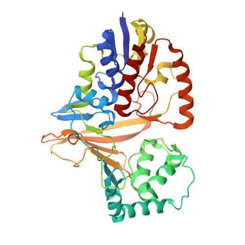
    1I8T

  • PubMed Abstract: 

    Uridine diphosphogalactofuranose (UDP-Galf ) is the precursor of the d-galactofuranose (Galf ) residues found in bacterial and parasitic cell walls, including those of many pathogens, such as Mycobacterium tuberculosis and Trypanosoma cruzi. UDP-Galf is made from UDP-galactopyranose (UDP-Galp) by the enzyme UDP-galactopyranose mutase (mutase). The mutase enzyme is essential for the viability of mycobacteria and is not found in humans, making it a viable therapeutic target. The mechanism by which mutase achieves the unprecedented ring contraction of a nonreducing sugar is unclear. We have solved the crystal structure of Escherichia coli mutase to 2.4 A resolution. The novel structure shows that the flavin nucleotide is located in a cleft lined with conserved residues. Site-directed mutagenesis studies indicate that this cleft contains the active site, with the sugar ring of the substrate UDP-galactose adjacent to the exposed isoalloxazine ring of FAD. Assay results establish that the enzyme is active only when flavin is reduced. We conclude that mutase most likely functions by transient reduction of substrate.


  • Organizational Affiliation

    The Centre for Biomolecular Sciences, The University, St. Andrews, Scotland KY16 9ST, UK.


Macromolecules
Find similar proteins by:  (by identity cutoff)  |  3D Structure
Entity ID: 1
MoleculeChains Sequence LengthOrganismDetailsImage
UDP-GALACTOPYRANOSE MUTASE
A, B
367Escherichia coliMutation(s): 0 
EC: 5.4.99.9
UniProt
Find proteins for P37747 (Escherichia coli (strain K12))
Explore P37747 
Go to UniProtKB:  P37747
Entity Groups  
Sequence Clusters30% Identity50% Identity70% Identity90% Identity95% Identity100% Identity
UniProt GroupP37747
Sequence Annotations
Expand
  • Reference Sequence
Small Molecules
Ligands 1 Unique
IDChains Name / Formula / InChI Key2D Diagram3D Interactions
FAD
Query on FAD

Download Ideal Coordinates CCD File 
C [auth A],
D [auth B]
FLAVIN-ADENINE DINUCLEOTIDE
C27 H33 N9 O15 P2
VWWQXMAJTJZDQX-UYBVJOGSSA-N
Experimental Data & Validation

Experimental Data

  • Method: X-RAY DIFFRACTION
  • Resolution: 2.40 Å
  • R-Value Free: 0.245 
  • R-Value Work: 0.185 
  • Space Group: P 21 21 21
Unit Cell:
Length ( Å )Angle ( ˚ )
a = 56.642α = 90
b = 98.123β = 90
c = 132.476γ = 90
Software Package:
Software NamePurpose
COMPLEXmodel building
CNSrefinement
MOSFLMdata reduction
CCP4data scaling
COMPLEXphasing

Structure Validation

View Full Validation Report


Ligand Structure Quality Assessment 


Entry History 

Deposition Data

Revision History  (Full details and data files)

  • Version 1.0: 2001-10-03
    Type: Initial release
  • Version 1.1: 2008-04-27
    Changes: Version format compliance
  • Version 1.2: 2011-07-13
    Changes: Version format compliance
  • Version 1.3: 2014-11-12
    Changes: Structure summary
  • Version 1.4: 2017-10-04
    Changes: Advisory
  • Version 1.5: 2020-09-02
    Changes: Derived calculations, Structure summary
  • Version 1.6: 2024-02-07
    Changes: Advisory, Data collection, Database references