1D8S

ESCHERICHIA COLI F1 ATPASE


Experimental Data Snapshot

  • Method: X-RAY DIFFRACTION
  • Resolution: 4.40 Å

wwPDB Validation   3D Report Full Report


This is version 1.4 of the entry. See complete history


Literature

Structural features of the gamma subunit of the Escherichia coli F(1) ATPase revealed by a 4.4-A resolution map obtained by x-ray crystallography.

Hausrath, A.C.Gruber, G.Matthews, B.W.Capaldi, R.A.

(1999) Proc Natl Acad Sci U S A 96: 13697-13702

  • DOI: https://doi.org/10.1073/pnas.96.24.13697
  • Primary Citation of Related Structures:  
    1D8S

  • PubMed Abstract: 

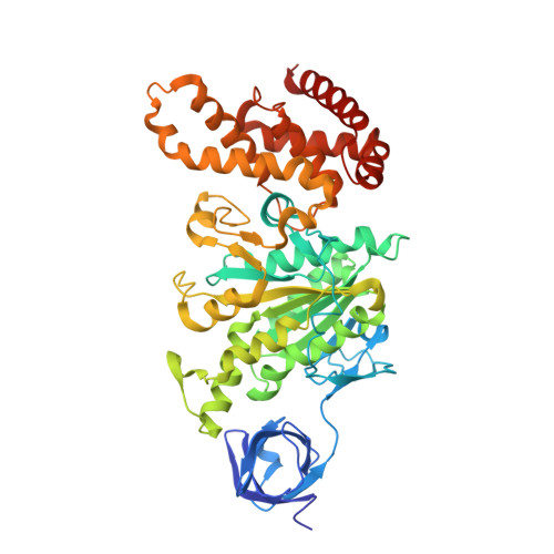
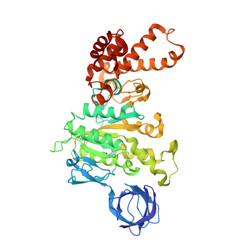
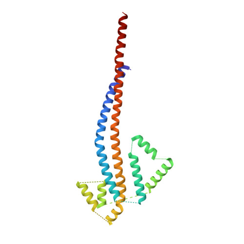
    The F(1) part of the F(1)F(O) ATP synthase from Escherichia coli has been crystallized and its structure determined to 4.4-A resolution by using molecular replacement based on the structure of the beef-heart mitochondrial enzyme. The bacterial F(1) consists of five subunits with stoichiometry alpha(3), beta(3), gamma, delta, and epsilon. delta was removed before crystallization. In agreement with the structure of the beef-heart mitochondrial enzyme, although not that from rat liver, the present study suggests that the alpha and beta subunits are arranged in a hexagonal barrel but depart from exact 3-fold symmetry. In the structures of both beef heart and rat-liver mitochondrial F(1), less than half of the structure of the gamma subunit was seen because of presumed disorder in the crystals. The present electron-density map includes a number of rod-shaped features which appear to correspond to additional alpha-helical regions within the gamma subunit. These suggest that the gamma subunit traverses the full length of the stalk that links the F(1) and F(O) parts and makes significant contacts with the c subunit ring of F(O).


  • Organizational Affiliation

    Institute of Molecular Biology, Howard Hughes Medical Institute, Department of Physics, 1229 University of Oregon, Eugene, OR 97403-1229, USA.


Macromolecules
Find similar proteins by:  (by identity cutoff)  |  3D Structure
Entity ID: 1
MoleculeChains Sequence LengthOrganismDetailsImage
F1 ATPASE (ALPHA SUBUNIT)
A, B, C
492Escherichia coliMutation(s): 0 
Sequence Annotations
Expand
  • Reference Sequence
Find similar proteins by:  (by identity cutoff)  |  3D Structure
Entity ID: 2
MoleculeChains Sequence LengthOrganismDetailsImage
F1 ATPASE (BETA SUBUNIT)
D, E, F
467Escherichia coliMutation(s): 0 
Sequence Annotations
Expand
  • Reference Sequence
Find similar proteins by:  (by identity cutoff)  |  3D Structure
Entity ID: 3
MoleculeChains Sequence LengthOrganismDetailsImage
F1 ATPASE (GAMMA SUBUNIT)214Escherichia coliMutation(s): 0 
Sequence Annotations
Expand
  • Reference Sequence
Experimental Data & Validation

Experimental Data

  • Method: X-RAY DIFFRACTION
  • Resolution: 4.40 Å
  • Space Group: C 2 2 21
Unit Cell:
Length ( Å )Angle ( ˚ )
a = 180.68α = 90
b = 197.52β = 90
c = 237.9γ = 90
Software Package:
Software NamePurpose
AMoREphasing
TNTrefinement
MOSFLMdata reduction
SCALAdata scaling

Structure Validation

View Full Validation Report


Entry History 

Deposition Data

Revision History  (Full details and data files)

  • Version 1.0: 1999-12-03
    Type: Initial release
  • Version 1.1: 2008-04-27
    Changes: Version format compliance
  • Version 1.2: 2011-07-13
    Changes: Version format compliance
  • Version 1.3: 2020-07-22
    Changes: Data collection, Other, Refinement description
  • Version 1.4: 2024-02-07
    Changes: Database references