8SOS

Human CD1d presenting sphingomyelin C24:1 in complex with VHH nanobody 1D17


Experimental Data Snapshot

  • Method: X-RAY DIFFRACTION
  • Resolution: 2.33 Å
  • R-Value Free: 0.230 
  • R-Value Work: 0.209 
  • R-Value Observed: 0.210 

wwPDB Validation   3D Report Full Report


Ligand Structure Quality Assessment 


This is version 1.0 of the entry. See complete history


Literature

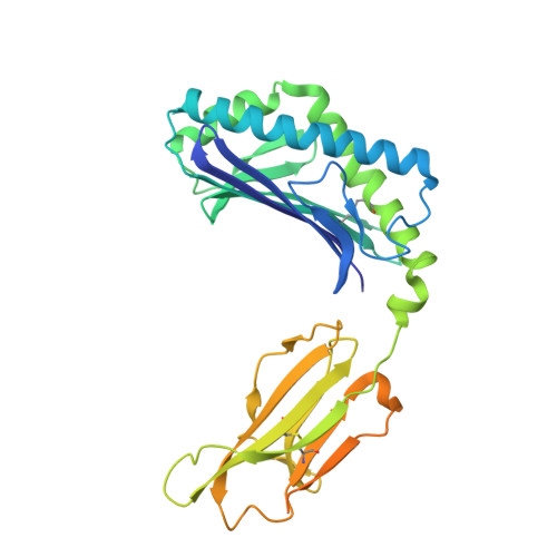
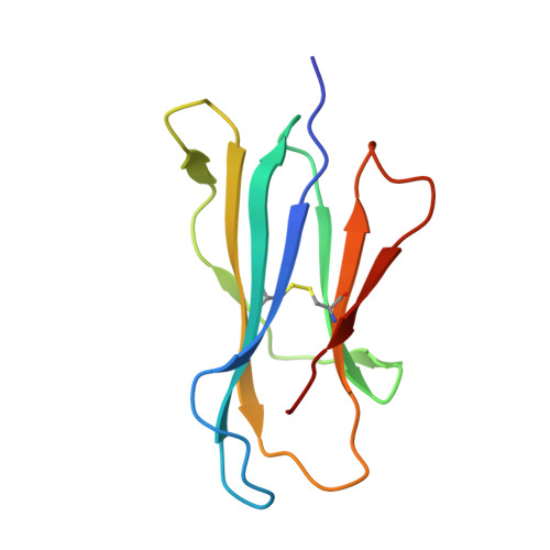
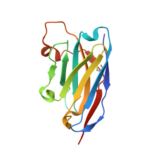
Enhanced CD1d phosphatidylserine presentation using a single-domain antibody promotes immunomodulatory CD1d-TIM-3 interactions.

Lameris, R.Shahine, A.Veth, M.Westerman, B.Godfrey, D.I.Lutje Hulsik, D.Brouwer, P.Rossjohn, J.de Gruijl, T.D.van der Vliet, H.J.

(2023) J Immunother Cancer 11

  • DOI: https://doi.org/10.1136/jitc-2023-007631
  • Primary Citation of Related Structures:  
    8SOS

  • PubMed Abstract: 

    CD1d is a monomorphic major histocompatibility complex class I-like molecule that presents lipid antigens to distinct T-cell subsets and can be expressed by various malignancies. Antibody-mediated targeting of CD1d on multiple myeloma cells was reported to induce apoptosis and could therefore constitute a novel therapeutic approach. To determine how a CD1d-specific single-domain antibody (VHH) enhances binding of the early apoptosis marker annexin V to CD1d + tumor cells we use in vitro cell-based assays and CRISPR-Cas9-mediated gene editing, and to determine the structure of the VHH1D17-CD1d(endogenous lipid) complex we use X-ray crystallography. Anti-CD1d VHH1D17 strongly enhances annexin V binding to CD1d + tumor cells but this does not reflect induction of apoptosis. Instead, we show that VHH1D17 enhances presentation of phosphatidylserine (PS) in CD1d and that this is saposin dependent. The crystal structure of the VHH1D17-CD1d(endogenous lipid) complex demonstrates that VHH1D17 binds the A'-pocket of CD1d, leaving the lipid headgroup solvent exposed, and has an electro-negatively charged patch which could be involved in the enhanced PS presentation by CD1d. Presentation of PS in CD1d does not trigger phagocytosis but leads to greatly enhanced binding of T-cell immunoglobulin and mucin domain containing molecules (TIM)-1 to TIM-3, TIM-4 and induces TIM-3 signaling. Our findings reveal the existence of an immune modulatory CD1d(PS)-TIM axis with potentially unexpected implications for immune regulation in both physiological and pathological conditions.


  • Organizational Affiliation

    Department of Medical Oncology, Cancer Center Amsterdam, Amsterdam UMC Location VUmc, Amsterdam, The Netherlands.


Macromolecules
Find similar proteins by:  (by identity cutoff)  |  3D Structure
Entity ID: 1
MoleculeChains Sequence LengthOrganismDetailsImage
Antigen-presenting glycoprotein CD1dA,
D [auth E]
347Homo sapiensMutation(s): 0 
Gene Names: CD1D
UniProt & NIH Common Fund Data Resources
Find proteins for P05412 (Homo sapiens)
Explore P05412 
Go to UniProtKB:  P05412
PHAROS:  P05412
GTEx:  ENSG00000177606 
Find proteins for P15813 (Homo sapiens)
Explore P15813 
Go to UniProtKB:  P15813
PHAROS:  P15813
GTEx:  ENSG00000158473 
Entity Groups  
Sequence Clusters30% Identity50% Identity70% Identity90% Identity95% Identity100% Identity
UniProt GroupsP15813P05412
Sequence Annotations
Expand
  • Reference Sequence
Find similar proteins by:  (by identity cutoff)  |  3D Structure
Entity ID: 2
MoleculeChains Sequence LengthOrganismDetailsImage
Beta-2-microglobulinB,
E [auth F]
100Homo sapiensMutation(s): 0 
Gene Names: B2MCDABP0092HDCMA22P
UniProt & NIH Common Fund Data Resources
Find proteins for P61769 (Homo sapiens)
Explore P61769 
Go to UniProtKB:  P61769
PHAROS:  P61769
GTEx:  ENSG00000166710 
Entity Groups  
Sequence Clusters30% Identity50% Identity70% Identity90% Identity95% Identity100% Identity
UniProt GroupP61769
Sequence Annotations
Expand
  • Reference Sequence
Find similar proteins by:  (by identity cutoff)  |  3D Structure
Entity ID: 3
MoleculeChains Sequence LengthOrganismDetailsImage
Nanobody VHH ID17C [auth D],
F [auth G]
127Lama glamaMutation(s): 0 
Entity Groups  
Sequence Clusters30% Identity50% Identity70% Identity90% Identity95% Identity100% Identity
Sequence Annotations
Expand
  • Reference Sequence
Experimental Data & Validation

Experimental Data

  • Method: X-RAY DIFFRACTION
  • Resolution: 2.33 Å
  • R-Value Free: 0.230 
  • R-Value Work: 0.209 
  • R-Value Observed: 0.210 
  • Space Group: P 32 2 1
Unit Cell:
Length ( Å )Angle ( ˚ )
a = 136.449α = 90
b = 136.449β = 90
c = 166.767γ = 120
Software Package:
Software NamePurpose
PHENIXrefinement
XDSdata reduction
Aimlessdata scaling
PHASERphasing

Structure Validation

View Full Validation Report


Ligand Structure Quality Assessment 


Entry History & Funding Information

Deposition Data


Funding OrganizationLocationGrant Number
Australian Research Council (ARC)AustraliaDE210101031

Revision History  (Full details and data files)

  • Version 1.0: 2023-12-13
    Type: Initial release