8BLT

Structure of Lactobacillus salivarius (Ls) bile salt hydrolase(BSH) in complex with taurocholate (TCA)


Experimental Data Snapshot

  • Method: X-RAY DIFFRACTION
  • Resolution: 2.10 Å
  • R-Value Free: 0.213 
  • R-Value Work: 0.180 
  • R-Value Observed: 0.182 

wwPDB Validation   3D Report Full Report


Ligand Structure Quality Assessment 


This is version 1.3 of the entry. See complete history


Literature

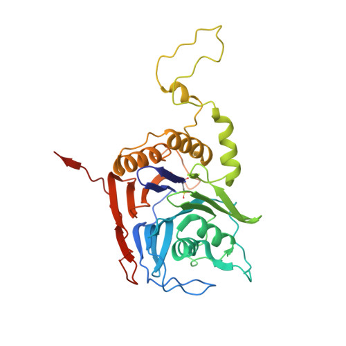
Characterization of the mechanism of bile salt hydrolase substrate specificity by experimental and computational analyses.

Karlov, D.S.Long, S.L.Zeng, X.Xu, F.Lal, K.Cao, L.Hayoun, K.Lin, J.Joyce, S.A.Tikhonova, I.G.

(2023) Structure 31: 629

  • DOI: https://doi.org/10.1016/j.str.2023.02.014
  • Primary Citation of Related Structures:  
    8BLS, 8BLT

  • PubMed Abstract: 

    Bile salt hydrolases (BSHs) are currently being investigated as target enzymes for metabolic regulators in humans and as growth promoters in farm animals. Understanding structural features underlying substrate specificity is necessary for inhibitor design. Here, we used a multidisciplinary workflow including mass spectrometry, mutagenesis, molecular dynamic simulations, machine learning, and crystallography to demonstrate substrate specificity in Lactobacillus salivarius BSH, the most abundant enzyme in human and farm animal intestines. We show the preference of substrates with a taurine head and a dehydroxylated sterol ring for hydrolysis. A regression model that correlates the relative rates of hydrolysis of various substrates in various enzyme mutants with the residue-substrate interaction energies guided the identification of structural determinants of substrate binding and specificity. In addition, we found T208 from another BSH protomer regulating the hydrolysis. The designed workflow can be used for fast and comprehensive characterization of enzymes with a broad range of substrates.


  • Organizational Affiliation

    School of Pharmacy, Medical Biology Centre, Queen's University Belfast, BT9 7BL Northern Ireland, UK.


Macromolecules
Find similar proteins by:  (by identity cutoff)  |  3D Structure
Entity ID: 1
MoleculeChains Sequence LengthOrganismDetailsImage
Bile salt hydrolase
A, B, C, D, E
A, B, C, D, E, F, G, H
325Ligilactobacillus salivariusMutation(s): 0 
UniProt
Find proteins for J7H3P9 (Ligilactobacillus salivarius)
Explore J7H3P9 
Go to UniProtKB:  J7H3P9
Entity Groups  
Sequence Clusters30% Identity50% Identity70% Identity90% Identity95% Identity100% Identity
UniProt GroupJ7H3P9
Sequence Annotations
Expand
  • Reference Sequence
Experimental Data & Validation

Experimental Data

  • Method: X-RAY DIFFRACTION
  • Resolution: 2.10 Å
  • R-Value Free: 0.213 
  • R-Value Work: 0.180 
  • R-Value Observed: 0.182 
  • Space Group: P 1 21 1
Unit Cell:
Length ( Å )Angle ( ˚ )
a = 84.759α = 90
b = 94.605β = 90.26
c = 168.215γ = 90
Software Package:
Software NamePurpose
REFMACrefinement
Aimlessdata scaling
iMOSFLMdata reduction
PHASERphasing
PDB_EXTRACTdata extraction

Structure Validation

View Full Validation Report


Ligand Structure Quality Assessment 


Entry History & Funding Information

Deposition Data


Funding OrganizationLocationGrant Number
National Institute of Food and Agriculture (NIFA, United States)United States2018-67015-27475
Other government05935-NAGpro
Other governmentDAFM 17-RD-US-ROI
Other governmentKJCX20220422
Engineering and Physical Sciences Research CouncilUnited KingdomEP/R029407/1 and EP/W03204X/1

Revision History  (Full details and data files)

  • Version 1.0: 2023-03-08
    Type: Initial release
  • Version 1.1: 2023-04-05
    Changes: Database references
  • Version 1.2: 2023-05-17
    Changes: Database references
  • Version 1.3: 2024-02-07
    Changes: Data collection, Refinement description