5FT2

Sub-tomogram averaging of Lassa virus glycoprotein spike from virus- like particles at pH 5


Experimental Data Snapshot

  • Method: ELECTRON MICROSCOPY
  • Resolution: 16.4 Å
  • Aggregation State: PARTICLE 
  • Reconstruction Method: TOMOGRAPHY 

wwPDB Validation   3D Report Full Report


This is version 2.0 of the entry. See complete history


Literature

Acidic Ph-Induced Conformations and Lamp1 Binding of the Lassa Virus Glycoprotein Spike.

Li, S.Sun, Z.Pryce, R.Parsy, M.Fehling, S.K.Schlie, K.Siebert, C.A.Garten, W.Bowden, T.A.Strecker, T.Huiskonen, J.T.

(2016) PLoS Pathog 12: 5418

  • DOI: https://doi.org/10.1371/journal.ppat.1005418
  • Primary Citation of Related Structures:  
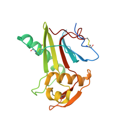
    5FT2

  • PubMed Abstract: 

    Lassa virus is an enveloped, bi-segmented RNA virus and the most prevalent and fatal of all Old World arenaviruses. Virus entry into the host cell is mediated by a tripartite surface spike complex, which is composed of two viral glycoprotein subunits, GP1 and GP2, and the stable signal peptide. Of these, GP1 binds to cellular receptors and GP2 catalyzes fusion between the viral envelope and the host cell membrane during endocytosis. The molecular structure of the spike and conformational rearrangements induced by low pH, prior to fusion, remain poorly understood. Here, we analyzed the three-dimensional ultrastructure of Lassa virus using electron cryotomography. Sub-tomogram averaging yielded a structure of the glycoprotein spike at 14-Å resolution. The spikes are trimeric, cover the virion envelope, and connect to the underlying matrix. Structural changes to the spike, following acidification, support a viral entry mechanism dependent on binding to the lysosome-resident receptor LAMP1 and further dissociation of the membrane-distal GP1 subunits.


  • Organizational Affiliation

    Division of Structural Biology, Wellcome Trust Centre for Human Genetics, University of Oxford, Oxford, United Kingdom.


Macromolecules
Find similar proteins by:  (by identity cutoff)  |  3D Structure
Entity ID: 1
MoleculeChains Sequence LengthOrganismDetailsImage
PRE-GLYCOPROTEIN POLYPROTEIN GP COMPLEXA [auth B]172Lassa virus JosiahMutation(s): 0 
UniProt
Find proteins for P08669 (Lassa virus (strain Mouse/Sierra Leone/Josiah/1976))
Explore P08669 
Go to UniProtKB:  P08669
Entity Groups  
Sequence Clusters30% Identity50% Identity70% Identity90% Identity95% Identity100% Identity
UniProt GroupP08669
Sequence Annotations
Expand
  • Reference Sequence
Oligosaccharides

Help

Entity ID: 2
MoleculeChains Length2D Diagram Glycosylation3D Interactions
2-acetamido-2-deoxy-beta-D-glucopyranose-(1-4)-2-acetamido-2-deoxy-beta-D-glucopyranoseB [auth A],
C
2N-Glycosylation
Glycosylation Resources
GlyTouCan:  G42666HT
GlyCosmos:  G42666HT
GlyGen:  G42666HT
Small Molecules
Ligands 1 Unique
IDChains Name / Formula / InChI Key2D Diagram3D Interactions
NAG
Query on NAG

Download Ideal Coordinates CCD File 
D [auth B],
E [auth B]
2-acetamido-2-deoxy-beta-D-glucopyranose
C8 H15 N O6
OVRNDRQMDRJTHS-FMDGEEDCSA-N
Experimental Data & Validation

Experimental Data

  • Method: ELECTRON MICROSCOPY
  • Resolution: 16.4 Å
  • Aggregation State: PARTICLE 
  • Reconstruction Method: TOMOGRAPHY 
EM Software:
TaskSoftware PackageVersion
RECONSTRUCTIONDynamo
RECONSTRUCTIONIMOD

Structure Validation

View Full Validation Report


Entry History 

Deposition Data

Revision History  (Full details and data files)

  • Version 1.0: 2016-03-02
    Type: Initial release
  • Version 1.1: 2017-08-02
    Changes: Data collection, Refinement description
  • Version 1.2: 2017-08-30
    Changes: Data collection
  • Version 2.0: 2020-07-29
    Type: Remediation
    Reason: Carbohydrate remediation
    Changes: Atomic model, Data collection, Derived calculations, Structure summary