


Structure of EAV NSP11 K170A mutant at 3.19A
Zhang, M.F., Chen, Z.Z.To be published.
Experimental Data Snapshot
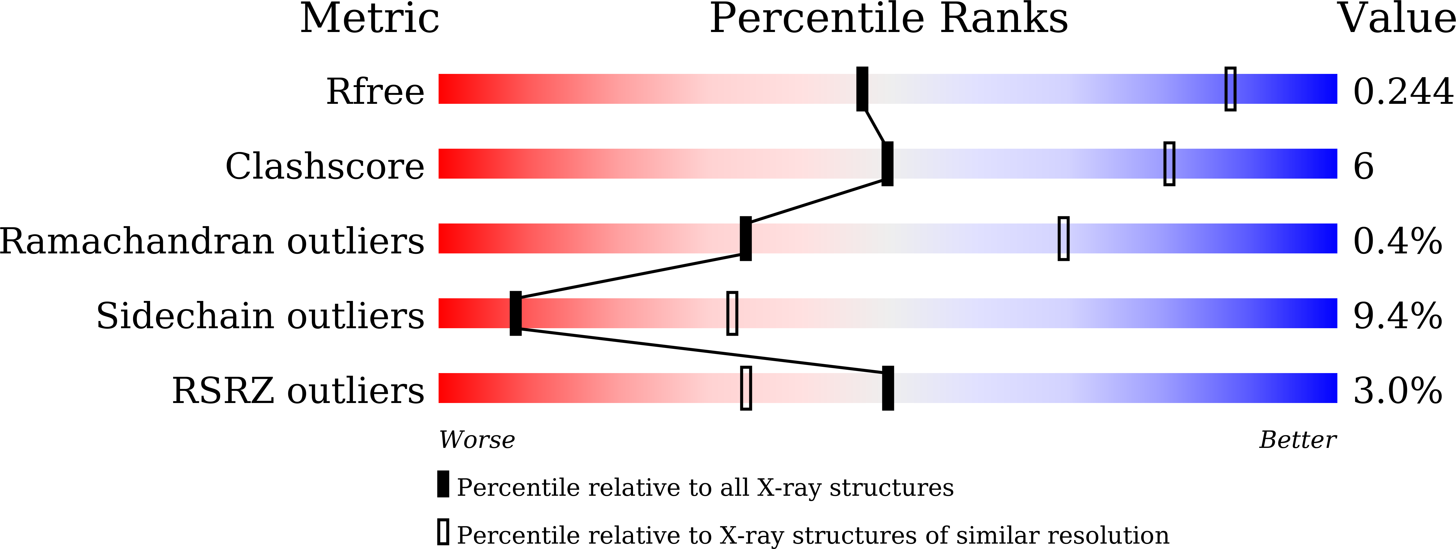
wwPDB Validation 3D Report Full Report
Entity ID: 1 | |||||
|---|---|---|---|---|---|
| Molecule | Chains | Sequence Length | Organism | Details | Image |
| Non-structural protein 11 | 219 | Equine arteritis virus Bucyrus | Mutation(s): 1 Gene Names: rep, 1a-1b | 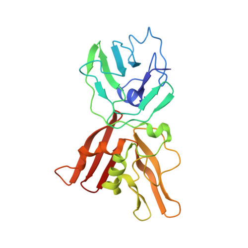 | |
UniProt | |||||
Find proteins for P19811 (Equine arteritis virus (strain Bucyrus)) Explore P19811 Go to UniProtKB: P19811 | |||||
Entity Groups | |||||
| Sequence Clusters | 30% Identity50% Identity70% Identity90% Identity95% Identity100% Identity | ||||
| UniProt Group | P19811 | ||||
Sequence AnnotationsExpand | |||||
| |||||
| Length ( Å ) | Angle ( ˚ ) |
|---|---|
| a = 249.184 | α = 90 |
| b = 249.184 | β = 90 |
| c = 226.899 | γ = 120 |
| Software Name | Purpose |
|---|---|
| PHENIX | refinement |
| HKL-2000 | data reduction |
| HKL-2000 | data scaling |
| BALBES | phasing |
RCSB PDB (citation) is hosted by
RCSB PDB is a member of the

