4XC5

CRYSTAL STRUCTURE OF THE T1L REOVIRUS ATTACHMENT PROTEIN SIGMA1


Experimental Data Snapshot

  • Method: X-RAY DIFFRACTION
  • Resolution: 2.20 Å
  • R-Value Free: 0.223 
  • R-Value Work: 0.190 
  • R-Value Observed: 0.193 

wwPDB Validation   3D Report Full Report


This is version 2.0 of the entry. See complete history


Literature

Structure of Serotype 1 Reovirus Attachment Protein sigma 1 in Complex with Junctional Adhesion Molecule A Reveals a Conserved Serotype-Independent Binding Epitope.

Stettner, E.Dietrich, M.H.Reiss, K.Dermody, T.S.Stehle, T.

(2015) J Virol 89: 6136-6140

  • DOI: https://doi.org/10.1128/JVI.00433-15
  • Primary Citation of Related Structures:  
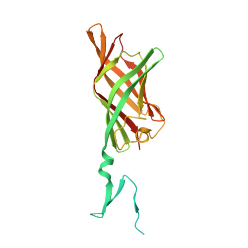
    4XC5

  • PubMed Abstract: 

    Mammalian orthoreoviruses use glycans and junctional adhesion molecule A (JAM-A) as attachment receptors. We determined the structure of serotype 1 reovirus attachment protein σ1 alone and in complex with JAM-A. Comparison with the structure of serotype 3 reovirus σ1 bound to JAM-A reveals that both σ1 proteins engage JAM-A with similar affinities and via conserved binding epitopes. Thus, σ1-JAM-A interactions are unlikely to explain the differences in pathogenesis displayed by these reovirus serotypes.


  • Organizational Affiliation

    Interfaculty Institute of Biochemistry, University of Tübingen, Tübingen, Germany.


Macromolecules
Find similar proteins by:  (by identity cutoff)  |  3D Structure
Entity ID: 1
MoleculeChains Sequence LengthOrganismDetailsImage
Outer capsid protein sigma-1
A, B, C
218Mammalian orthoreovirus 1 LangMutation(s): 0 
Gene Names: S1
UniProt
Find proteins for P04506 (Reovirus type 1 (strain Lang))
Explore P04506 
Go to UniProtKB:  P04506
Entity Groups  
Sequence Clusters30% Identity50% Identity70% Identity90% Identity95% Identity100% Identity
UniProt GroupP04506
Sequence Annotations
Expand
  • Reference Sequence
Small Molecules
Ligands 4 Unique
IDChains Name / Formula / InChI Key2D Diagram3D Interactions
GOL
Query on GOL

Download Ideal Coordinates CCD File 
G [auth A]
H [auth A]
L [auth B]
M [auth B]
N [auth B]
G [auth A],
H [auth A],
L [auth B],
M [auth B],
N [auth B],
R [auth C],
S [auth C]
GLYCEROL
C3 H8 O3
PEDCQBHIVMGVHV-UHFFFAOYSA-N
ACT
Query on ACT

Download Ideal Coordinates CCD File 
F [auth A],
K [auth B],
Q [auth C]
ACETATE ION
C2 H3 O2
QTBSBXVTEAMEQO-UHFFFAOYSA-M
CL
Query on CL

Download Ideal Coordinates CCD File 
D [auth A],
I [auth B],
O [auth C]
CHLORIDE ION
Cl
VEXZGXHMUGYJMC-UHFFFAOYSA-M
MG
Query on MG

Download Ideal Coordinates CCD File 
E [auth A],
J [auth B],
P [auth C]
MAGNESIUM ION
Mg
JLVVSXFLKOJNIY-UHFFFAOYSA-N
Experimental Data & Validation

Experimental Data

  • Method: X-RAY DIFFRACTION
  • Resolution: 2.20 Å
  • R-Value Free: 0.223 
  • R-Value Work: 0.190 
  • R-Value Observed: 0.193 
  • Space Group: I 21 21 21
Unit Cell:
Length ( Å )Angle ( ˚ )
a = 112.93α = 90
b = 112.96β = 90
c = 113.22γ = 90
Software Package:
Software NamePurpose
BUSTERrefinement
XDSdata reduction
XSCALEdata scaling
PHASERphasing

Structure Validation

View Full Validation Report


Entry History & Funding Information

Deposition Data


Funding OrganizationLocationGrant Number
National Institutes of HealthUnited StatesR01 AI076983

Revision History  (Full details and data files)

  • Version 1.0: 2015-04-01
    Type: Initial release
  • Version 1.1: 2015-04-08
    Changes: Database references
  • Version 1.2: 2015-05-13
    Changes: Database references
  • Version 1.3: 2016-07-13
    Changes: Data collection
  • Version 2.0: 2024-01-10
    Changes: Atomic model, Author supporting evidence, Data collection, Database references, Derived calculations, Refinement description