4X13

JC Polyomavirus genotype 3 VP1 in complex with LSTc pentasaccharide


Experimental Data Snapshot

  • Method: X-RAY DIFFRACTION
  • Resolution: 2.00 Å
  • R-Value Free: 0.178 
  • R-Value Work: 0.157 
  • R-Value Observed: 0.158 

wwPDB Validation   3D Report Full Report


This is version 3.1 of the entry. See complete history


Literature

The Greater Affinity of JC Polyomavirus Capsid for alpha 2,6-Linked Lactoseries Tetrasaccharide c than for Other Sialylated Glycans Is a Major Determinant of Infectivity.

Stroh, L.J.Maginnis, M.S.Blaum, B.S.Nelson, C.D.Neu, U.Gee, G.V.O'Hara, B.A.Motamedi, N.DiMaio, D.Atwood, W.J.Stehle, T.

(2015) J Virol 89: 6364-6375

  • DOI: https://doi.org/10.1128/JVI.00489-15
  • Primary Citation of Related Structures:  
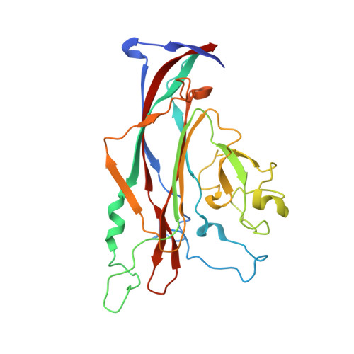
    4X0Y, 4X0Z, 4X10, 4X11, 4X12, 4X13, 4X14, 4X15, 4X16, 4X17

  • PubMed Abstract: 

    The human JC polyomavirus (JCPyV) establishes an asymptomatic, persistent infection in the kidneys of the majority of the population and is the causative agent of the fatal demyelinating disease progressive multifocal leukoencephalopathy (PML) in immunosuppressed individuals. The Mad-1 strain of JCPyV, a brain isolate, was shown earlier to require α2,6-linked sialic acid on the lactoseries tetrasaccharide c (LSTc) glycan for attachment to host cells. In contrast, a JCPyV kidney isolate type 3 strain, WT3, has been reported to interact with sialic acid-containing gangliosides, but the role of these glycans in JCPyV infection has remained unclear. To help rationalize these findings and probe the effects of strain-specific differences on receptor binding, we performed a comprehensive analysis of the glycan receptor specificities of these two representative JCPyV strains using high-resolution X-ray crystallography and nuclear magnetic resonance (NMR) spectroscopy, and correlated these data with the results of infectivity assays. We show here that capsid proteins of Mad-1 and WT3 JCPyV can both engage LSTc as well as multiple sialylated gangliosides. However, the binding affinities exhibit subtle differences, with the highest affinity observed for LSTc. Engagement of LSTc is a prerequisite for functional receptor engagement, while the more weakly binding gangliosides are not required for productive infection. Our findings highlight the complexity of virus-carbohydrate interactions and demonstrate that subtle differences in binding affinities, rather than the binding event alone, help determine tissue tropism and viral pathogenesis. Viral infection is initiated by attachment to receptors on host cells, and this event plays an important role in viral disease. We investigated the receptor-binding properties of human JC polyomavirus (JCPyV), a virus that resides in the kidneys of the majority of the population and can cause the fatal demyelinating disease progressive multifocal leukoencephalopathy (PML) in the brains of immunosuppressed individuals. JCPyV has been reported to interact with multiple carbohydrate receptors, and we sought to clarify how the interactions between JCPyV and cellular carbohydrate receptors influenced infection. Here we demonstrate that JCPyV can engage numerous sialylated carbohydrate receptors. However, the virus displays preferential binding to LSTc, and only LSTc mediates a productive infection. Our findings demonstrate that subtle differences in binding affinity, rather than receptor engagement alone, are a key determinant of viral infection.


  • Organizational Affiliation

    Interfaculty Institute of Biochemistry, University of Tübingen, Tübingen, Germany.


Macromolecules
Find similar proteins by:  (by identity cutoff)  |  3D Structure
Entity ID: 1
MoleculeChains Sequence LengthOrganismDetailsImage
Major capsid protein
A, B, C, D, E
272JC polyomavirus type 3Mutation(s): 0 
Gene Names: VP1
UniProt
Find proteins for P90498 (JC polyomavirus)
Explore P90498 
Go to UniProtKB:  P90498
Entity Groups  
Sequence Clusters30% Identity50% Identity70% Identity90% Identity95% Identity100% Identity
UniProt GroupP90498
Sequence Annotations
Expand
  • Reference Sequence
Oligosaccharides

Help

Entity ID: 2
MoleculeChains Length2D Diagram Glycosylation3D Interactions
N-acetyl-alpha-neuraminic acid-(2-6)-beta-D-galactopyranose-(1-4)-2-acetamido-2-deoxy-beta-D-glucopyranose-(1-3)-beta-D-galactopyranose-(1-4)-beta-D-glucopyranose
F, G
5N/A
Glycosylation Resources
GlyTouCan:  G83513YI
GlyCosmos:  G83513YI
GlyGen:  G83513YI
Entity ID: 3
MoleculeChains Length2D Diagram Glycosylation3D Interactions
N-acetyl-alpha-neuraminic acid-(2-6)-beta-D-galactopyranose-(1-4)-2-acetamido-2-deoxy-beta-D-glucopyranose-(1-3)-beta-D-galactopyranose
H
4N/A
Glycosylation Resources
GlyTouCan:  G03509EL
GlyCosmos:  G03509EL
GlyGen:  G03509EL
Small Molecules
Ligands 3 Unique
IDChains Name / Formula / InChI Key2D Diagram3D Interactions
GOL
Query on GOL

Download Ideal Coordinates CCD File 
AA [auth C]
CA [auth D]
DA [auth D]
FA [auth D]
HA [auth E]
AA [auth C],
CA [auth D],
DA [auth D],
FA [auth D],
HA [auth E],
I [auth A],
M [auth A],
T [auth B],
U [auth B],
X [auth C],
Z [auth C]
GLYCEROL
C3 H8 O3
PEDCQBHIVMGVHV-UHFFFAOYSA-N
EDO
Query on EDO

Download Ideal Coordinates CCD File 
EA [auth D]
IA [auth E]
J [auth A]
JA [auth E]
K [auth A]
EA [auth D],
IA [auth E],
J [auth A],
JA [auth E],
K [auth A],
KA [auth E],
L [auth A],
LA [auth E],
O [auth B],
P [auth B],
Q [auth B],
R [auth B],
S [auth B],
V [auth B],
Y [auth C]
1,2-ETHANEDIOL
C2 H6 O2
LYCAIKOWRPUZTN-UHFFFAOYSA-N
K
Query on K

Download Ideal Coordinates CCD File 
BA [auth C],
GA [auth D],
MA [auth E],
N [auth A],
W [auth B]
POTASSIUM ION
K
NPYPAHLBTDXSSS-UHFFFAOYSA-N
Experimental Data & Validation

Experimental Data

  • Method: X-RAY DIFFRACTION
  • Resolution: 2.00 Å
  • R-Value Free: 0.178 
  • R-Value Work: 0.157 
  • R-Value Observed: 0.158 
  • Space Group: C 1 2 1
Unit Cell:
Length ( Å )Angle ( ˚ )
a = 150.12α = 90
b = 96.28β = 109.81
c = 128.14γ = 90
Software Package:
Software NamePurpose
REFMACrefinement
Cootmodel building
PHASERphasing
XDSdata reduction

Structure Validation

View Full Validation Report


Entry History & Funding Information

Deposition Data


Funding OrganizationLocationGrant Number
National Institutes of HealthUnited States--

Revision History  (Full details and data files)

  • Version 1.0: 2015-04-22
    Type: Initial release
  • Version 1.1: 2015-05-27
    Changes: Database references
  • Version 2.0: 2018-01-31
    Changes: Atomic model, Author supporting evidence, Derived calculations
  • Version 3.0: 2020-07-29
    Type: Remediation
    Reason: Carbohydrate remediation
    Changes: Atomic model, Data collection, Derived calculations, Structure summary
  • Version 3.1: 2024-01-10
    Changes: Data collection, Database references, Derived calculations, Refinement description, Structure summary