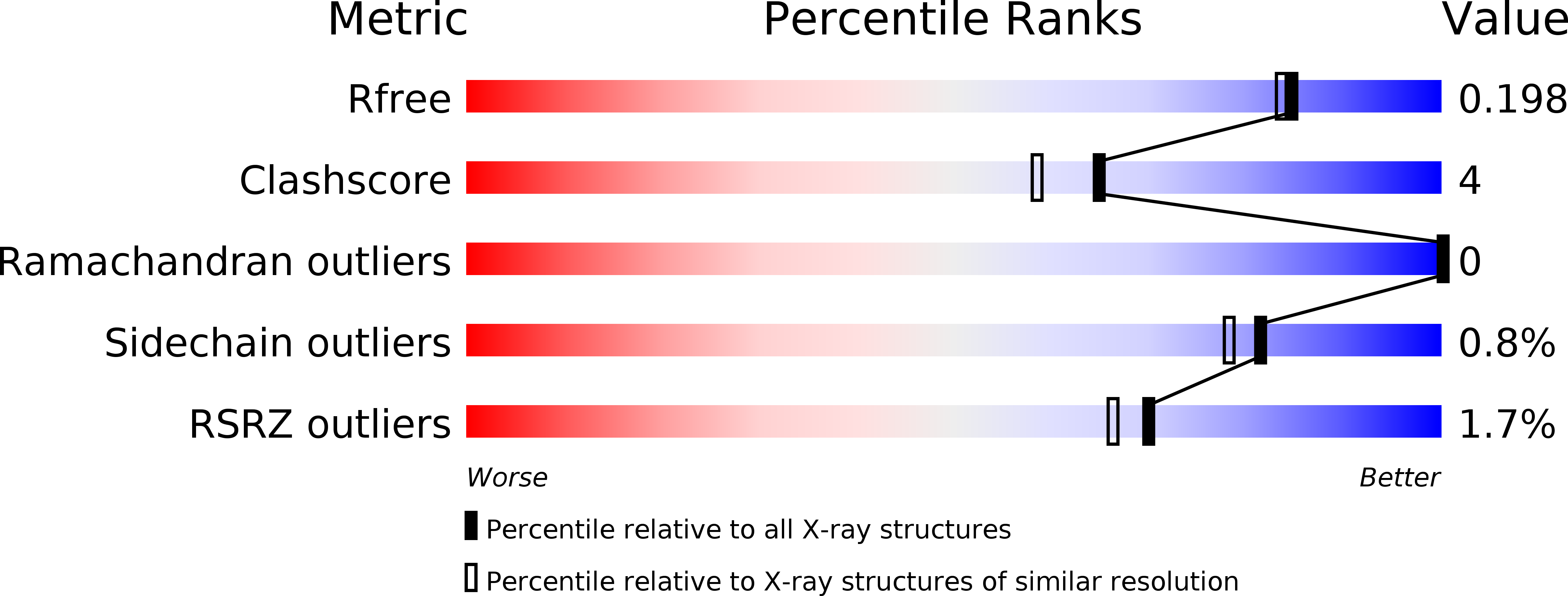
An open conformation determined by a structural switch for 2A protease from coxsackievirus A16.
Sun, Y., Wang, X., Yuan, S., Dang, M., Li, X., Zhang, X.C., Rao, Z.(2013) Protein Cell 4: 782-792
- PubMed: 24026848
- DOI: https://doi.org/10.1007/s13238-013-3914-z
- Primary Citation of Related Structures:
4MG3 - PubMed Abstract:
Coxsackievirus A16 belongs to the family Picornaviridae, and is a major agent of hand-foot-and-mouth disease that infects mostly children, and to date no vaccines or antiviral therapies are available. 2A protease of enterovirus is a nonstructural protein and possesses both self-cleavage activity and the ability to cleave the eukaryotic translation initiation factor 4G. Here we present the crystal structure of coxsackievirus A16 2A protease, which interestingly forms hexamers in crystal as well as in solution. This structure shows an open conformation, with its active site accessible, ready for substrate binding and cleavage activity. In conjunction with a previously reported "closed" state structure of human rhinovirus 2, we were able to develop a detailed hypothesis for the conformational conversion triggered by two "switcher" residues Glu88 and Tyr89 located within the bll2-cII loop. Substrate recognition assays revealed that amino acid residues P1', P2 and P4 are essential for substrate specificity, which was verified by our substrate binding model. In addition, we compared the in vitro cleavage efficiency of 2A proteases from coxsackievirus A16 and enterovirus 71 upon the same substrates by fluorescence resonance energy transfer (FRET), and observed higher protease activity of enterovirus 71 compared to that of coxsackievirus A16. In conclusion, our study shows an open conformation of coxsackievirus A16 2A protease and the underlying mechanisms for conformational conversion and substrate specificity. These new insights should facilitate the future rational design of efficient 2A protease inhibitors.
Organizational Affiliation:
National Laboratory of Biomacromolecules, Institute of Biophysics, Chinese Academy of Sciences, Beijing, 100101, China.


