Structural investigation of a viral ortholog of human NEIL2/3 DNA glycosylases.
Prakash, A., Eckenroth, B.E., Averill, A.M., Imamura, K., Wallace, S.S., Doublie, S.(2013) DNA Repair (Amst) 12: 1062-1071
- PubMed: 24120312
- DOI: https://doi.org/10.1016/j.dnarep.2013.09.004
- Primary Citation of Related Structures:
4MB7 - PubMed Abstract:
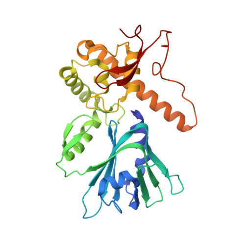
Assault to DNA that leads to oxidative base damage is repaired by the base excision repair (BER) pathway with specialized enzymes called DNA glycosylases catalyzing the first step of this pathway. These glycosylases can be categorized into two families: the HhH superfamily, which includes endonuclease III (or Nth), and the Fpg/Nei family, which comprises formamidopyrimidine DNA glycosylase (or Fpg) and endonuclease VIII (or Nei). In humans there are three Nei-like (NEIL) glycosylases: NEIL1, 2, and 3. Here we present the first crystal structure of a viral ortholog of the human NEIL2/NEIL3 proteins, Mimivirus Nei2 (MvNei2), determined at 2.04Å resolution. The C-terminal region of the MvNei2 enzyme comprises two conserved DNA binding motifs: the helix-two-turns-helix (H2TH) motif and a C-H-C-C type zinc-finger similar to that of human NEIL2. The N-terminal region of MvNei2 is most closely related to NEIL3. Like NEIL3, MvNei2 bears a valine at position 2 instead of the usual proline and it lacks two of the three conserved void-filling residues present in other members of the Fpg/Nei family. Mutational analysis of the only conserved void-filling residue methionine 72 to alanine yields an MvNei2 variant with impaired glycosylase activity. Mutation of the adjacent His73 causes the enzyme to be more productive thereby suggesting a plausible role for this residue in the DNA lesion search process.
Organizational Affiliation:
Department of Microbiology and Molecular Genetics, The Markey Center for Molecular Genetics, University of Vermont, Stafford Hall, 95 Carrigan Drive, Burlington, VT 05405-0068, United States.