4DGR

Influenza Subtype 9 Neuraminidase Benzoic Acid Inhibitor Complex


Experimental Data Snapshot

  • Method: X-RAY DIFFRACTION
  • Resolution: 1.55 Å
  • R-Value Free: 0.152 
  • R-Value Work: 0.121 
  • R-Value Observed: 0.123 

wwPDB Validation   3D Report Full Report


Ligand Structure Quality Assessment 


This is version 2.1 of the entry. See complete history


Literature

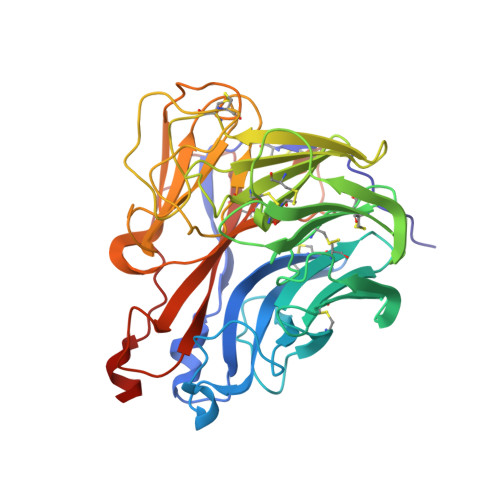
Crystal structure of a new benzoic acid inhibitor of influenza neuraminidase bound with a new tilt induced by overpacking sub-site C6.

Venkatramani, L.Johnson, E.S.Kolavi, G.Air, G.M.Brouillette, W.J.Mooers, B.H.

(2012) BMC Struct Biol 12: 7-7

  • DOI: https://doi.org/10.1186/1472-6807-12-7
  • Primary Citation of Related Structures:  
    4DGR

  • PubMed Abstract: 

    Influenza neuraminidase (NA) is an important target for antiviral inhibitors since its active site is highly conserved such that inhibitors can be cross-reactive against multiple types and subtypes of influenza. Here, we discuss the crystal structure of neuraminidase subtype N9 complexed with a new benzoic acid based inhibitor (2) that was designed to add contacts by overpacking one side of the active site pocket. Inhibitor 2 uses benzoic acid to mimic the pyranose ring, a bis-(hydroxymethyl)-substituted 2-pyrrolidinone ring in place of the N-acetyl group of the sialic acid, and a branched aliphatic structure to fill the sialic acid C6 subsite. Inhibitor 2 {4-[2,2-bis(hydroxymethyl)-5-oxo-pyrrolidin-1-yl]-3-[(dipropylamino)methyl)]benzoic acid} was soaked into crystals of neuraminidase of A/tern/Australia/G70c/75 (N9), and the structure refined with 1.55 Å X-ray data. The benzene ring of the inhibitor tilted 8.9° compared to the previous compound (1), and the number of contacts, including hydrogen bonds, increased. However, the IC50 for compound 2 remained in the low micromolar range, likely because one propyl group was disordered. In this high-resolution structure of NA isolated from virus grown in chicken eggs, we found electron density for additional sugar units on the N-linked glycans compared to previous neuraminidase structures. In particular, seven mannoses and two N-acetylglucosamines are visible in the glycan attached to Asn200. This long, branched high-mannose glycan makes significant contacts with the neighboring subunit. We designed inhibitor 2 with an extended substituent at C4-corresponding to C6 of sialic acid-to increase the contact surface in the C6-subsite and to force the benzene ring to tilt to maximize these interactions while retaining the interactions of the carboxylate and the pyrolidinone substituents. The crystal structure at 1.55 Å showed that we partially succeeded in that the ring in 2 is tilted relative to 1 and the number of contacts increased, but one hydrophobic branch makes no contacts, perhaps explaining why the IC50 did not decrease. Future design efforts will include branches of unequal length so that both branches may be accommodated in the C6-subsite without conformational disorder. The high-mannose glycan attached to Asn200 makes several inter-subunit contacts and appears to stabilize the tetramer.


  • Organizational Affiliation

    Department of Biochemistry and Molecular Biology, University of Oklahoma Health Sciences Center, 941 Stanton L, Young Blvd, Oklahoma City, OK 73104, USA.


Macromolecules
Find similar proteins by:  (by identity cutoff)  |  3D Structure
Entity ID: 1
MoleculeChains Sequence LengthOrganismDetailsImage
Neuraminidase389Influenza A virus (A/tern/Australia/G70C/1975(H11N9))Mutation(s): 0 
EC: 3.2.1.18
UniProt
Find proteins for P03472 (Influenza A virus (strain A/Tern/Australia/G70C/1975 H11N9))
Explore P03472 
Go to UniProtKB:  P03472
Entity Groups  
Sequence Clusters30% Identity50% Identity70% Identity90% Identity95% Identity100% Identity
UniProt GroupP03472
Sequence Annotations
Expand
  • Reference Sequence
Oligosaccharides

Help

Entity ID: 2
MoleculeChains Length2D Diagram Glycosylation3D Interactions
alpha-D-mannopyranose-(1-2)-alpha-D-mannopyranose-(1-2)-alpha-D-mannopyranose-(1-3)-[alpha-D-mannopyranose-(1-3)-[alpha-D-mannopyranose-(1-6)]alpha-D-mannopyranose-(1-6)]beta-D-mannopyranose-(1-4)-2-acetamido-2-deoxy-beta-D-glucopyranose-(1-4)-2-acetamido-2-deoxy-beta-D-glucopyranose
B
9N-Glycosylation
Glycosylation Resources
GlyTouCan:  G68668TB
GlyCosmos:  G68668TB
GlyGen:  G68668TB
Entity ID: 3
MoleculeChains Length2D Diagram Glycosylation3D Interactions
2-acetamido-2-deoxy-beta-D-glucopyranose-(1-4)-2-acetamido-2-deoxy-beta-D-glucopyranose
C
2N-Glycosylation
Glycosylation Resources
GlyTouCan:  G42666HT
GlyCosmos:  G42666HT
GlyGen:  G42666HT
Entity ID: 4
MoleculeChains Length2D Diagram Glycosylation3D Interactions
beta-D-mannopyranose-(1-4)-2-acetamido-2-deoxy-beta-D-glucopyranose-(1-4)-2-acetamido-2-deoxy-beta-D-glucopyranose
D
3N-Glycosylation
Glycosylation Resources
GlyTouCan:  G15407YE
GlyCosmos:  G15407YE
GlyGen:  G15407YE
Small Molecules
Ligands 6 Unique
IDChains Name / Formula / InChI Key2D Diagram3D Interactions
3LV
Query on 3LV

Download Ideal Coordinates CCD File 
I [auth A]4-[2,2-bis(hydroxymethyl)-5-oxopyrrolidin-1-yl]-3-[(dipropylamino)methyl]benzoic acid
C20 H30 N2 O5
IYODICUSAICAQL-UHFFFAOYSA-N
GLC
Query on GLC

Download Ideal Coordinates CCD File 
F [auth A]alpha-D-glucopyranose
C6 H12 O6
WQZGKKKJIJFFOK-DVKNGEFBSA-N
BGC
Query on BGC

Download Ideal Coordinates CCD File 
E [auth A],
G [auth A],
H [auth A]
beta-D-glucopyranose
C6 H12 O6
WQZGKKKJIJFFOK-VFUOTHLCSA-N
PO4
Query on PO4

Download Ideal Coordinates CCD File 
J [auth A]PHOSPHATE ION
O4 P
NBIIXXVUZAFLBC-UHFFFAOYSA-K
CA
Query on CA

Download Ideal Coordinates CCD File 
K [auth A],
L [auth A]
CALCIUM ION
Ca
BHPQYMZQTOCNFJ-UHFFFAOYSA-N
K
Query on K

Download Ideal Coordinates CCD File 
M [auth A]POTASSIUM ION
K
NPYPAHLBTDXSSS-UHFFFAOYSA-N
Binding Affinity Annotations 
IDSourceBinding Affinity
3LV PDBBind:  4DGR IC50: 9100 (nM) from 1 assay(s)
Binding MOAD:  4DGR IC50: 9100 (nM) from 1 assay(s)
Experimental Data & Validation

Experimental Data

  • Method: X-RAY DIFFRACTION
  • Resolution: 1.55 Å
  • R-Value Free: 0.152 
  • R-Value Work: 0.121 
  • R-Value Observed: 0.123 
  • Space Group: I 4 3 2
Unit Cell:
Length ( Å )Angle ( ˚ )
a = 180.969α = 90
b = 180.969β = 90
c = 180.969γ = 90
Software Package:
Software NamePurpose
Blu-Icedata collection
PHENIXrefinement
XDSdata reduction
SCALAdata scaling

Structure Validation

View Full Validation Report


Ligand Structure Quality Assessment 


Entry History 

Deposition Data

Revision History  (Full details and data files)

  • Version 1.0: 2012-05-16
    Type: Initial release
  • Version 1.1: 2012-06-20
    Changes: Database references
  • Version 1.2: 2017-11-15
    Changes: Advisory, Refinement description
  • Version 2.0: 2020-07-29
    Type: Remediation
    Reason: Carbohydrate remediation
    Changes: Atomic model, Data collection, Derived calculations, Structure summary
  • Version 2.1: 2023-09-13
    Changes: Advisory, Data collection, Database references, Refinement description, Structure summary