3V8X

The crystal structure of transferrin binding protein A (TbpA) from Neisserial meningitidis serogroup B in complex with full length human transferrin


Experimental Data Snapshot

  • Method: X-RAY DIFFRACTION
  • Resolution: 2.60 Å
  • R-Value Free: 0.267 
  • R-Value Work: 0.208 
  • R-Value Observed: 0.211 

wwPDB Validation   3D Report Full Report


Ligand Structure Quality Assessment 


This is version 2.0 of the entry. See complete history


Literature

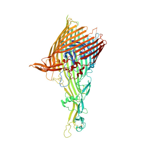
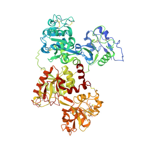
Structural basis for iron piracy by pathogenic Neisseria.

Noinaj, N.Easley, N.C.Oke, M.Mizuno, N.Gumbart, J.Boura, E.Steere, A.N.Zak, O.Aisen, P.Tajkhorshid, E.Evans, R.W.Gorringe, A.R.Mason, A.B.Steven, A.C.Buchanan, S.K.

(2012) Nature 483: 53-58

  • DOI: https://doi.org/10.1038/nature10823
  • Primary Citation of Related Structures:  
    3SKP, 3V83, 3V89, 3V8U, 3V8X

  • PubMed Abstract: 

    Neisseria are obligate human pathogens causing bacterial meningitis, septicaemia and gonorrhoea. Neisseria require iron for survival and can extract it directly from human transferrin for transport across the outer membrane. The transport system consists of TbpA, an integral outer membrane protein, and TbpB, a co-receptor attached to the cell surface; both proteins are potentially important vaccine and therapeutic targets. Two key questions driving Neisseria research are how human transferrin is specifically targeted, and how the bacteria liberate iron from transferrin at neutral pH. To address these questions, we solved crystal structures of the TbpA-transferrin complex and of the corresponding co-receptor TbpB. We characterized the TbpB-transferrin complex by small-angle X-ray scattering and the TbpA-TbpB-transferrin complex by electron microscopy. Our studies provide a rational basis for the specificity of TbpA for human transferrin, show how TbpA promotes iron release from transferrin, and elucidate how TbpB facilitates this process.


  • Organizational Affiliation

    Laboratory of Molecular Biology, National Institute of Diabetes and Digestive and Kidney Diseases, US National Institutes of Health, Bethesda, Maryland 20892, USA.


Macromolecules
Find similar proteins by:  (by identity cutoff)  |  3D Structure
Entity ID: 1
MoleculeChains Sequence LengthOrganismDetailsImage
Transferrin-binding protein 1904Neisseria meningitidis serogroup BMutation(s): 2 
Gene Names: tbp1NMB0461
Membrane Entity: Yes 
UniProt
Find proteins for Q9K0U9 (Neisseria meningitidis serogroup B (strain MC58))
Explore Q9K0U9 
Go to UniProtKB:  Q9K0U9
Entity Groups  
Sequence Clusters30% Identity50% Identity70% Identity90% Identity95% Identity100% Identity
UniProt GroupQ9K0U9
Sequence Annotations
Expand
  • Reference Sequence
Find similar proteins by:  (by identity cutoff)  |  3D Structure
Entity ID: 2
MoleculeChains Sequence LengthOrganismDetailsImage
Serotransferrin698Homo sapiensMutation(s): 1 
Gene Names: TFPRO1400
Membrane Entity: Yes 
UniProt & NIH Common Fund Data Resources
Find proteins for P02787 (Homo sapiens)
Explore P02787 
Go to UniProtKB:  P02787
PHAROS:  P02787
GTEx:  ENSG00000091513 
Entity Groups  
Sequence Clusters30% Identity50% Identity70% Identity90% Identity95% Identity100% Identity
UniProt GroupP02787
Sequence Annotations
Expand
  • Reference Sequence
Oligosaccharides

Help

Entity ID: 3
MoleculeChains Length2D Diagram Glycosylation3D Interactions
N-acetyl-alpha-neuraminic acid-(2-6)-beta-D-galactopyranose-(1-4)-2-acetamido-2-deoxy-beta-D-glucopyranose-(1-4)-alpha-D-mannopyranose-(1-3)-[beta-D-galactopyranose-(1-4)-2-acetamido-2-deoxy-beta-D-glucopyranose-(1-4)-alpha-D-mannopyranose-(1-6)]beta-D-mannopyranose-(1-4)-2-acetamido-2-deoxy-beta-D-glucopyranose-(1-4)-2-acetamido-2-deoxy-beta-D-glucopyranose
C
10N-Glycosylation
Glycosylation Resources
GlyTouCan:  G98736SM
GlyCosmos:  G98736SM
GlyGen:  G98736SM
Entity ID: 4
MoleculeChains Length2D Diagram Glycosylation3D Interactions
N-acetyl-alpha-neuraminic acid-(2-6)-beta-D-galactopyranose-(1-4)-2-acetamido-2-deoxy-beta-D-glucopyranose-(1-4)-alpha-D-mannopyranose-(1-6)-[2-acetamido-2-deoxy-beta-D-glucopyranose-(1-4)-alpha-D-mannopyranose-(1-3)]beta-D-mannopyranose-(1-4)-2-acetamido-2-deoxy-beta-D-glucopyranose-(1-4)-2-acetamido-2-deoxy-beta-D-glucopyranose
D
9N-Glycosylation
Glycosylation Resources
GlyTouCan:  G74608QW
GlyCosmos:  G74608QW
GlyGen:  G74608QW
Small Molecules
Ligands 1 Unique
IDChains Name / Formula / InChI Key2D Diagram3D Interactions
C8E
Query on C8E

Download Ideal Coordinates CCD File 
E [auth A],
F [auth A]
(HYDROXYETHYLOXY)TRI(ETHYLOXY)OCTANE
C16 H34 O5
FEOZZFHAVXYAMB-UHFFFAOYSA-N
Experimental Data & Validation

Experimental Data

  • Method: X-RAY DIFFRACTION
  • Resolution: 2.60 Å
  • R-Value Free: 0.267 
  • R-Value Work: 0.208 
  • R-Value Observed: 0.211 
  • Space Group: P 21 21 21
Unit Cell:
Length ( Å )Angle ( ˚ )
a = 91.014α = 90
b = 129.362β = 90
c = 198.589γ = 90
Software Package:
Software NamePurpose
SERGUIdata collection
PHASERphasing
PHENIXrefinement
HKL-2000data reduction
HKL-2000data scaling

Structure Validation

View Full Validation Report


Ligand Structure Quality Assessment 


Entry History 

Deposition Data

Revision History  (Full details and data files)

  • Version 1.0: 2012-02-29
    Type: Initial release
  • Version 1.1: 2012-04-18
    Changes: Database references
  • Version 2.0: 2020-07-29
    Type: Remediation
    Reason: Carbohydrate remediation
    Changes: Advisory, Atomic model, Data collection, Database references, Derived calculations, Structure summary