3QR7

Crystal structure of the C-terminal fragment of the bacteriophage P2 membrane-piercing protein gpV


Experimental Data Snapshot

  • Method: X-RAY DIFFRACTION
  • Resolution: 0.94 Å
  • R-Value Free: 0.112 
  • R-Value Work: 0.102 
  • R-Value Observed: 0.102 

wwPDB Validation   3D Report Full Report


This is version 1.3 of the entry. See complete history


Literature

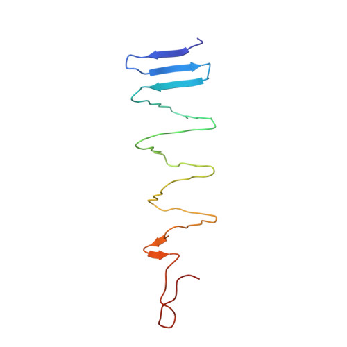
Phage pierces the host cell membrane with the iron-loaded spike.

Browning, C.Shneider, M.M.Bowman, V.D.Schwarzer, D.Leiman, P.G.

(2012) Structure 20: 326-339

  • DOI: https://doi.org/10.1016/j.str.2011.12.009
  • Primary Citation of Related Structures:  
    3PQH, 3PQI, 3QR7, 3QR8

  • PubMed Abstract: 

    Bacteriophages with contractile tails and the bacterial type VI secretion system have been proposed to use a special protein to create an opening in the host cell membrane during infection. These proteins have a modular architecture but invariably contain an oligonucleotide/oligosaccharide-binding (OB-fold) domain and a long β-helical C-terminal domain, which initiates the contact with the host cell membrane. Using X-ray crystallography and electron microscopy, we report the atomic structure of the membrane-piercing proteins from bacteriophages P2 and ϕ92 and identify the residues that constitute the membrane-attacking apex. Both proteins form compact spikes with a ∼10Å diameter tip that is stabilized by a centrally positioned iron ion bound by six histidine residues. The accumulated data strongly suggest that, in the process of membrane penetration, the spikes are translocated through the lipid bilayer without undergoing major unfolding.


  • Organizational Affiliation

    Institut de Physique des Systèmes Biologiques, Laboratory of Structural Biology and Biophysics, Ecole Polytechnique Fédérale de Lausanne (EPFL), BSP-415, CH-1015, Lausanne, Switzerland.


Macromolecules
Find similar proteins by:  (by identity cutoff)  |  3D Structure
Entity ID: 1
MoleculeChains Sequence LengthOrganismDetailsImage
Baseplate assembly protein V
A, B
115Peduovirus P2Mutation(s): 0 
Gene Names: V
UniProt
Find proteins for P31340 (Escherichia phage P2)
Explore P31340 
Go to UniProtKB:  P31340
Entity Groups  
Sequence Clusters30% Identity50% Identity70% Identity90% Identity95% Identity100% Identity
UniProt GroupP31340
Sequence Annotations
Expand
  • Reference Sequence
Small Molecules
Ligands 4 Unique
IDChains Name / Formula / InChI Key2D Diagram3D Interactions
FE
Query on FE

Download Ideal Coordinates CCD File 
E [auth A],
I [auth B]
FE (III) ION
Fe
VTLYFUHAOXGGBS-UHFFFAOYSA-N
CA
Query on CA

Download Ideal Coordinates CCD File 
D [auth A],
H [auth B]
CALCIUM ION
Ca
BHPQYMZQTOCNFJ-UHFFFAOYSA-N
CL
Query on CL

Download Ideal Coordinates CCD File 
C [auth A],
G [auth B]
CHLORIDE ION
Cl
VEXZGXHMUGYJMC-UHFFFAOYSA-M
NA
Query on NA

Download Ideal Coordinates CCD File 
F [auth A]SODIUM ION
Na
FKNQFGJONOIPTF-UHFFFAOYSA-N
Experimental Data & Validation

Experimental Data

  • Method: X-RAY DIFFRACTION
  • Resolution: 0.94 Å
  • R-Value Free: 0.112 
  • R-Value Work: 0.102 
  • R-Value Observed: 0.102 
  • Space Group: H 3 2
Unit Cell:
Length ( Å )Angle ( ˚ )
a = 49.069α = 90
b = 49.069β = 90
c = 463.896γ = 120
Software Package:
Software NamePurpose
RemDAqdata collection
SHELXmodel building
PHENIXrefinement
MOSFLMdata reduction
SCALAdata scaling
SHELXphasing

Structure Validation

View Full Validation Report


Entry History 

Deposition Data

Revision History  (Full details and data files)

  • Version 1.0: 2012-02-22
    Type: Initial release
  • Version 1.1: 2012-02-29
    Changes: Database references
  • Version 1.2: 2017-11-08
    Changes: Refinement description
  • Version 1.3: 2024-02-21
    Changes: Data collection, Database references, Derived calculations