3FVC

Crystal structure of a trimeric variant of the Epstein-Barr virus glycoprotein B


Experimental Data Snapshot

  • Method: X-RAY DIFFRACTION
  • Resolution: 3.20 Å
  • R-Value Free: 0.283 
  • R-Value Work: 0.242 
  • R-Value Observed: 0.246 

wwPDB Validation   3D Report Full Report


Ligand Structure Quality Assessment 


This is version 1.5 of the entry. See complete history


Literature

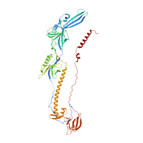
Structure of a trimeric variant of the Epstein-Barr virus glycoprotein B

Backovic, M.Longnecker, R.Jardetzky, T.S.

(2009) Proc Natl Acad Sci U S A 106: 2880-2885

  • DOI: https://doi.org/10.1073/pnas.0810530106
  • Primary Citation of Related Structures:  
    3FVC

  • PubMed Abstract: 

    Epstein-Barr virus (EBV) is a herpesvirus that is associated with development of malignancies of lymphoid tissue. EBV infections are life-long and occur in >90% of the population. Herpesviruses enter host cells in a process that involves fusion of viral and cellular membranes. The fusion apparatus is comprised of envelope glycoprotein B (gB) and a heterodimeric complex made of glycoproteins H and L. Glycoprotein B is the most conserved envelope glycoprotein in human herpesviruses, and the structure of gB from Herpes simplex virus 1 (HSV-1) is available. Here, we report the crystal structure of the secreted EBV gB ectodomain, which forms 16-nm long spike-like trimers, structurally homologous to the postfusion trimers of the fusion protein G of vesicular stomatitis virus (VSV). Comparative structural analyses of EBV gB and VSV G, which has been solved in its pre and postfusion states, shed light on gB residues that may be involved in conformational changes and membrane fusion. Also, the EBV gB structure reveals that, despite the high sequence conservation of gB in herpesviruses, the relative orientations of individual domains, the surface charge distributions, and the structural details of EBV gB differ from the HSV-1 protein, indicating regions and residues that may have important roles in virus-specific entry.


  • Organizational Affiliation

    Department of Biochemistry, Molecular Biology, and Cell Biology, Northwestern University, 2205 Tech Drive, Evanston, IL 60208.


Macromolecules
Find similar proteins by:  (by identity cutoff)  |  3D Structure
Entity ID: 1
MoleculeChains Sequence LengthOrganismDetailsImage
Glycoprotein GP110663Human herpesvirus 4 strain B95-8Mutation(s): 6 
Gene Names: BALF4VGLB_EBV
UniProt
Find proteins for P03188 (Epstein-Barr virus (strain B95-8))
Explore P03188 
Go to UniProtKB:  P03188
Entity Groups  
Sequence Clusters30% Identity50% Identity70% Identity90% Identity95% Identity100% Identity
UniProt GroupP03188
Sequence Annotations
Expand
  • Reference Sequence
Experimental Data & Validation

Experimental Data

  • Method: X-RAY DIFFRACTION
  • Resolution: 3.20 Å
  • R-Value Free: 0.283 
  • R-Value Work: 0.242 
  • R-Value Observed: 0.246 
  • Space Group: P 3 2 1
Unit Cell:
Length ( Å )Angle ( ˚ )
a = 106.8α = 90
b = 106.8β = 90
c = 210.75γ = 120
Software Package:
Software NamePurpose
DENZOdata reduction
SCALEPACKdata scaling
PHASERphasing
REFMACrefinement
PDB_EXTRACTdata extraction
MAR345dtbdata collection

Structure Validation

View Full Validation Report


Ligand Structure Quality Assessment 


Entry History 

Deposition Data

  • Released Date: 2009-03-17 
  • Deposition Author(s): Backovic, M.

Revision History  (Full details and data files)

  • Version 1.0: 2009-03-17
    Type: Initial release
  • Version 1.1: 2011-07-13
    Changes: Advisory, Version format compliance
  • Version 1.2: 2017-11-01
    Changes: Refinement description
  • Version 1.3: 2020-07-29
    Type: Remediation
    Reason: Carbohydrate remediation
    Changes: Data collection, Derived calculations, Structure summary
  • Version 1.4: 2021-10-20
    Changes: Database references, Structure summary
  • Version 1.5: 2023-09-06
    Changes: Data collection, Refinement description