3FSN

Crystal structure of RPE65 at 2.14 angstrom resolution


Experimental Data Snapshot

  • Method: X-RAY DIFFRACTION
  • Resolution: 2.14 Å
  • R-Value Free: 0.216 
  • R-Value Work: 0.180 
  • R-Value Observed: 0.182 

wwPDB Validation   3D Report Full Report


This is version 1.3 of the entry. See complete history


Literature

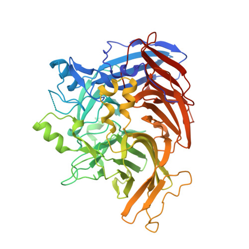
Crystal structure of native RPE65, the retinoid isomerase of the visual cycle.

Kiser, P.D.Golczak, M.Lodowski, D.T.Chance, M.R.Palczewski, K.

(2009) Proc Natl Acad Sci U S A 106: 17325-17330

  • DOI: https://doi.org/10.1073/pnas.0906600106
  • Primary Citation of Related Structures:  
    3FSN

  • PubMed Abstract: 

    Vertebrate vision is maintained by the retinoid (visual) cycle, a complex enzymatic pathway that operates in the retina to regenerate the visual chromophore, 11-cis-retinal. A key enzyme in this pathway is the microsomal membrane protein RPE65. This enzyme catalyzes the conversion of all-trans-retinyl esters to 11-cis-retinol in the retinal pigment epithelium (RPE). Mutations in RPE65 are known to be responsible for a subset of cases of the most common form of childhood blindness, Leber congenital amaurosis (LCA). Although retinoid isomerase activity has been attributed to RPE65, its catalytic mechanism remains a matter of debate. Also, the manner in which RPE65 binds to membranes and extracts retinoid substrates is unclear. To gain insight into these questions, we determined the crystal structure of native bovine RPE65 at 2.14-A resolution. The structural, biophysical, and biochemical data presented here provide the framework needed for an in-depth understanding of the mechanism of catalytic isomerization and membrane association, in addition to the role mutations that cause LCA have in disrupting protein function.


  • Organizational Affiliation

    Department of Pharmacology, School of Medicine, Case Western Reserve University, Cleveland, OH 44106-4965, USA.


Macromolecules
Find similar proteins by:  (by identity cutoff)  |  3D Structure
Entity ID: 1
MoleculeChains Sequence LengthOrganismDetailsImage
Retinal pigment epithelium-specific 65 kDa protein
A, B
533Bos taurusMutation(s): 1 
EC: 5.2.1.7
Membrane Entity: Yes 
UniProt
Find proteins for Q28175 (Bos taurus)
Explore Q28175 
Go to UniProtKB:  Q28175
Entity Groups  
Sequence Clusters30% Identity50% Identity70% Identity90% Identity95% Identity100% Identity
UniProt GroupQ28175
Sequence Annotations
Expand
  • Reference Sequence
Experimental Data & Validation

Experimental Data

  • Method: X-RAY DIFFRACTION
  • Resolution: 2.14 Å
  • R-Value Free: 0.216 
  • R-Value Work: 0.180 
  • R-Value Observed: 0.182 
  • Space Group: P 65
Unit Cell:
Length ( Å )Angle ( ˚ )
a = 176.516α = 90
b = 176.516β = 90
c = 86.862γ = 120
Software Package:
Software NamePurpose
HKL-2000data collection
SHELXSphasing
REFMACrefinement
HKL-2000data reduction
HKL-2000data scaling

Structure Validation

View Full Validation Report


Entry History 

Deposition Data

Revision History  (Full details and data files)

  • Version 1.0: 2009-09-29
    Type: Initial release
  • Version 1.1: 2011-07-13
    Changes: Advisory, Version format compliance
  • Version 1.2: 2018-01-24
    Changes: Structure summary
  • Version 1.3: 2024-02-21
    Changes: Data collection, Database references, Derived calculations, Refinement description