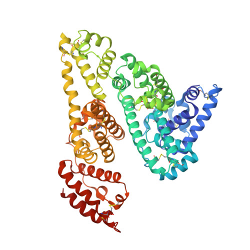
Crystallographic Analysis Reveals the Structural Basis of the High-Affinity Binding of Iophenoxic Acid to Human Serum Albumin.
Ryan, A.J., Chung, C.W., Curry, S.(2011) BMC Struct Biol 11: 18
- PubMed: 21501503
- DOI: https://doi.org/10.1186/1472-6807-11-18
- Primary Citation of Related Structures:
2YDF - PubMed Abstract:
Iophenoxic acid is an iodinated radiocontrast agent that was withdrawn from clinical use because of its exceptionally long half-life in the body, which was due in part to its high-affinity binding to human serum albumin (HSA). It was replaced by Iopanoic acid, which has an amino rather than a hydroxyl group at position 3 on the iodinated benzyl ring and, as a result, binds to albumin with lower affinity and is excreted more rapidly from the body. To understand how iophenoxic acid binds so tightly to albumin, we wanted to examine the structural basis of its interaction with HSA. We have determined the co-crystal structure of HSA in complex with iophenoxic acid at 2.75 Å resolution, revealing a total of four binding sites, two of which--in drugs sites 1 and 2 on the protein--are likely to be occupied at clinical doses. High-affinity binding of iophenoxic acid occurs at drug site 1. The structure reveals that polar and apolar groups on the compound are involved in its interactions with drug site 1. In particular, the 3-hydroxyl group makes three hydrogen bonds with the side-chains of Tyr 150 and Arg 257. The mode of binding to drug site 2 is similar except for the absence of a binding partner for the hydroxyl group on the benzyl ring of the compound. The HSA-iophenoxic acid structure indicates that high-affinity binding to drug site 1 is likely to be due to extensive desolvation of the compound, coupled with the ability of the binding pocket to provide a full set of salt-bridging or hydrogen bonding partners for its polar groups. Consistent with this interpretation, the structure also suggests that the lower-affinity binding of iopanoic acid arises because replacement of the 3-hydroxyl by an amino group eliminates hydrogen bonding to Arg 257. This finding underscores the importance of polar interactions in high-affinity binding to albumin.
Organizational Affiliation:
Biophysics Section, Blackett Laboratory, Imperial College, Exhibition Road, London, SW7 2AZ, UK.