2X9K

Structure of a E.coli porin


Experimental Data Snapshot

  • Method: X-RAY DIFFRACTION
  • Resolution: 2.18 Å
  • R-Value Free: 0.248 
  • R-Value Work: 0.213 
  • R-Value Observed: 0.215 

wwPDB Validation   3D Report Full Report


Ligand Structure Quality Assessment 


This is version 1.4 of the entry. See complete history


Literature

Correlation between the Ompg Secondary Structure and its Ph-Dependent Alterations Monitored by Ftir.

Korkmaz-Ozkan, F.Koster, S.Kuhlbrandt, W.Mantele, W.Yildiz, O.

(2010) J Mol Biol 401: 56

  • DOI: https://doi.org/10.1016/j.jmb.2010.06.015
  • Primary Citation of Related Structures:  
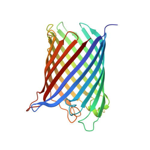
    2X9K

  • PubMed Abstract: 

    The channel activity of the outer-membrane protein G (OmpG) from Escherichia coli is pH-dependent. To investigate the role of the histidine pair His231/His261 in triggering channel opening and closing, we mutated both histidines to alanines and cysteines. Fourier transform infrared spectra revealed that the OmpG mutants stay-independent of pH-in an open conformation. Temperature ramp experiments indicate that the mutants are as stable as the open state of wild-type OmpG. The X-ray structure of the alanine-substituted OmpG mutant obtained at pH 6.5 confirms the constitutively open conformation. Compared to previous structures of the wild-type protein in the open and closed conformation, the mutant structure shows a difference in the extracellular loop L6 connecting beta-strands S12 and S13. A deletion of amino acids 220-228, which are thought to block the channel at low pH in wild-type OmpG, indicates conformational changes, which might be triggered by His231/His261.


  • Organizational Affiliation

    Institute of Biophysics, Goethe-University, Max-von-Laue-Str. 1, D-60438 Frankfurt am Main, Germany.


Macromolecules
Find similar proteins by:  (by identity cutoff)  |  3D Structure
Entity ID: 1
MoleculeChains Sequence LengthOrganismDetailsImage
OUTER MEMBRANE PROTEIN G280Escherichia coli K-12Mutation(s): 2 
Membrane Entity: Yes 
UniProt
Find proteins for P76045 (Escherichia coli (strain K12))
Explore P76045 
Go to UniProtKB:  P76045
Entity Groups  
Sequence Clusters30% Identity50% Identity70% Identity90% Identity95% Identity100% Identity
UniProt GroupP76045
Sequence Annotations
Expand
  • Reference Sequence
Experimental Data & Validation

Experimental Data

  • Method: X-RAY DIFFRACTION
  • Resolution: 2.18 Å
  • R-Value Free: 0.248 
  • R-Value Work: 0.213 
  • R-Value Observed: 0.215 
  • Space Group: P 21 21 21
Unit Cell:
Length ( Å )Angle ( ˚ )
a = 55.61α = 90
b = 70.96β = 90
c = 119.17γ = 90
Software Package:
Software NamePurpose
PHENIXrefinement
XDSdata reduction
XSCALEdata scaling

Structure Validation

View Full Validation Report


Ligand Structure Quality Assessment 


Entry History 

Deposition Data

Revision History  (Full details and data files)

  • Version 1.0: 2011-01-26
    Type: Initial release
  • Version 1.1: 2011-05-08
    Changes: Version format compliance
  • Version 1.2: 2011-07-13
    Changes: Version format compliance
  • Version 1.3: 2018-01-24
    Changes: Source and taxonomy
  • Version 1.4: 2020-07-29
    Type: Remediation
    Reason: Carbohydrate remediation
    Changes: Advisory, Data collection, Derived calculations, Other, Structure summary