2VWD

Nipah Virus Attachment Glycoprotein


Experimental Data Snapshot

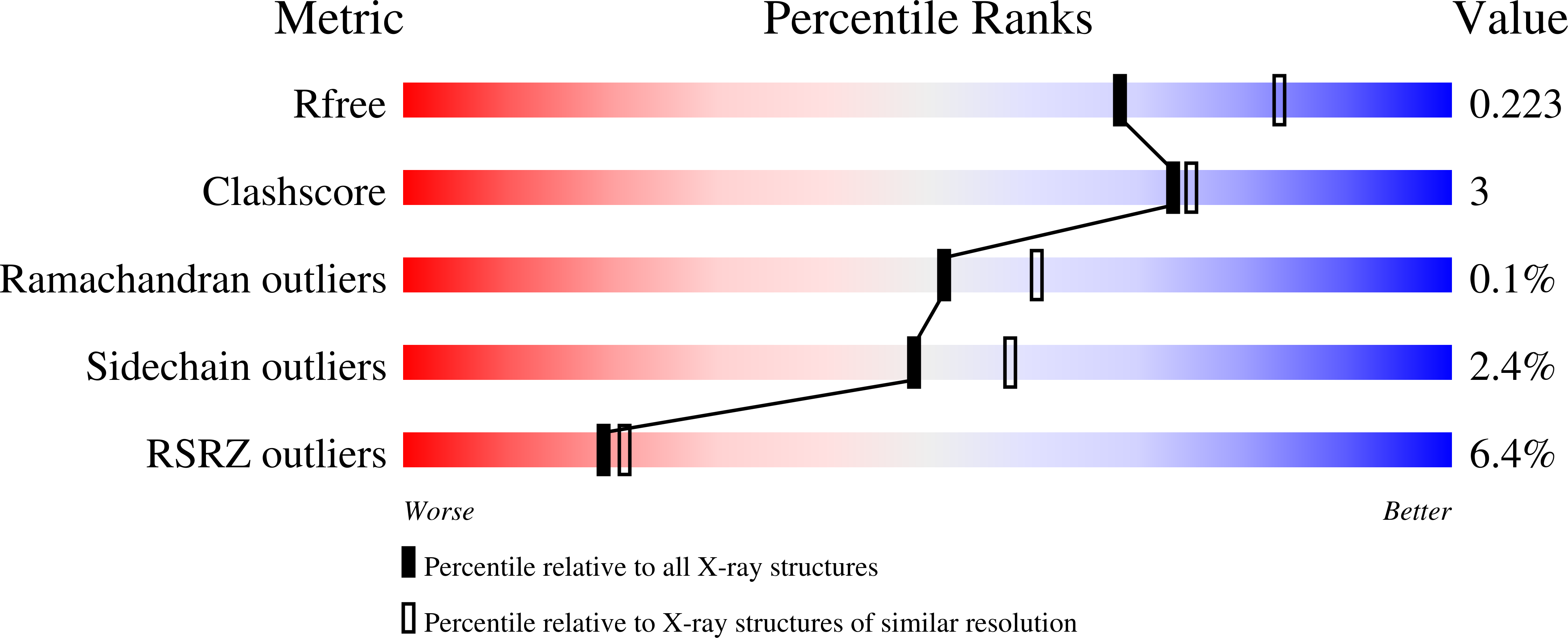
  • Method: X-RAY DIFFRACTION
  • Resolution: 2.25 Å
  • R-Value Free: 0.221 
  • R-Value Work: 0.172 
  • R-Value Observed: 0.175 

wwPDB Validation   3D Report Full Report


Ligand Structure Quality Assessment 


This is version 1.4 of the entry. See complete history


Literature

Crystal Structure and Carbohydrate Analysis of Nipah Virus Attachment Glycoprotein: A Template for Antiviral and Vaccine Design.

Bowden, T.A.Crispin, M.Harvey, D.J.Aricescu, A.R.Grimes, J.M.Jones, E.Y.Stuart, D.I.

(2008) J Virol 82: 11628

  • DOI: https://doi.org/10.1128/JVI.01344-08
  • Primary Citation of Related Structures:  
    2VWD

  • PubMed Abstract: 

    Two members of the paramyxovirus family, Nipah virus (NiV) and Hendra virus (HeV), are recent additions to a growing number of agents of emergent diseases which use bats as a natural host. Identification of ephrin-B2 and ephrin-B3 as cellular receptors for these viruses has enabled the development of immunotherapeutic reagents which prevent virus attachment and subsequent fusion. Here we present the structural analysis of the protein and carbohydrate components of the unbound viral attachment glycoprotein of NiV glycoprotein (NiV-G) at a 2.2-A resolution. Comparison with its ephrin-B2-bound form reveals that conformational changes within the envelope glycoprotein are required to achieve viral attachment. Structural differences are particularly pronounced in the 579-590 loop, a major component of the ephrin binding surface. In addition, the 236-245 loop is rather disordered in the unbound structure. We extend our structural characterization of NiV-G with mass spectrometric analysis of the carbohydrate moieties. We demonstrate that NiV-G is largely devoid of the oligomannose-type glycans that in viruses such as human immunodeficiency virus type 1 and Ebola virus influence viral tropism and the host immune response. Nevertheless, we find putative ligands for the endothelial cell lectin, LSECtin. Finally, by mapping structural conservation and glycosylation site positions from other members of the paramyxovirus family, we suggest the molecular surface involved in oligomerization. These results suggest possible pathways of virus-host interaction and strategies for the optimization of recombinant vaccines.


  • Organizational Affiliation

    Division of Structural Biology, University of Oxford, Henry Wellcome Building of Genomic Medicine, Roosevelt Drive, Oxford OX3 7BN, United Kingdom.


Macromolecules
Find similar proteins by:  (by identity cutoff)  |  3D Structure
Entity ID: 1
MoleculeChains Sequence LengthOrganismDetailsImage
HEMAGGLUTININ-NEURAMINIDASE
A, B
420Henipavirus nipahenseMutation(s): 0 
UniProt
Find proteins for Q9IH62 (Nipah virus)
Explore Q9IH62 
Go to UniProtKB:  Q9IH62
Entity Groups  
Sequence Clusters30% Identity50% Identity70% Identity90% Identity95% Identity100% Identity
UniProt GroupQ9IH62
Sequence Annotations
Expand
  • Reference Sequence
Small Molecules
Ligands 3 Unique
IDChains Name / Formula / InChI Key2D Diagram3D Interactions
NAG
Query on NAG

Download Ideal Coordinates CCD File 
I [auth A]
J [auth A]
K [auth A]
L [auth A]
M [auth A]
I [auth A],
J [auth A],
K [auth A],
L [auth A],
M [auth A],
R [auth B],
S [auth B],
T [auth B],
U [auth B],
V [auth B]
2-acetamido-2-deoxy-beta-D-glucopyranose
C8 H15 N O6
OVRNDRQMDRJTHS-FMDGEEDCSA-N
GBL
Query on GBL

Download Ideal Coordinates CCD File 
C [auth A]
D [auth A]
E [auth A]
F [auth A]
G [auth A]
C [auth A],
D [auth A],
E [auth A],
F [auth A],
G [auth A],
H [auth A],
P [auth B],
Q [auth B]
GAMMA-BUTYROLACTONE
C4 H6 O2
YEJRWHAVMIAJKC-UHFFFAOYSA-N
CL
Query on CL

Download Ideal Coordinates CCD File 
N [auth B],
O [auth B]
CHLORIDE ION
Cl
VEXZGXHMUGYJMC-UHFFFAOYSA-M
Experimental Data & Validation

Experimental Data

  • Method: X-RAY DIFFRACTION
  • Resolution: 2.25 Å
  • R-Value Free: 0.221 
  • R-Value Work: 0.172 
  • R-Value Observed: 0.175 
  • Space Group: P 1 21 1
Unit Cell:
Length ( Å )Angle ( ˚ )
a = 71.633α = 90
b = 86.936β = 108.32
c = 82.903γ = 90
Software Package:
Software NamePurpose
REFMACrefinement
HKL-2000data reduction
SCALEPACKdata scaling
PHASERphasing

Structure Validation

View Full Validation Report


Ligand Structure Quality Assessment 


Entry History 

Deposition Data

Revision History  (Full details and data files)

  • Version 1.0: 2008-10-07
    Type: Initial release
  • Version 1.1: 2011-07-13
    Changes: Advisory, Version format compliance
  • Version 1.2: 2018-02-28
    Changes: Source and taxonomy
  • Version 1.3: 2020-07-29
    Type: Remediation
    Reason: Carbohydrate remediation
    Changes: Data collection, Derived calculations, Other, Structure summary
  • Version 1.4: 2023-12-13
    Changes: Data collection, Database references, Refinement description, Structure summary