2VTU

crystal structure of bacteriophage MS2 covalent coat protein dimer


Experimental Data Snapshot

  • Method: X-RAY DIFFRACTION
  • Resolution: 3.50 Å
  • R-Value Free: 0.310 
  • R-Value Work: 0.272 
  • R-Value Observed: 0.272 

wwPDB Validation   3D Report Full Report


This is version 1.3 of the entry. See complete history


Literature

Crystal Packing of a Bacteriophage MS2 Coat Protein Mutant Corresponds to Octahedral Particles.

Plevka, P.Tars, K.Liljas, L.

(2008) Protein Sci 17: 1731

  • DOI: https://doi.org/10.1110/ps.036905.108
  • Primary Citation of Related Structures:  
    2VTU

  • PubMed Abstract: 

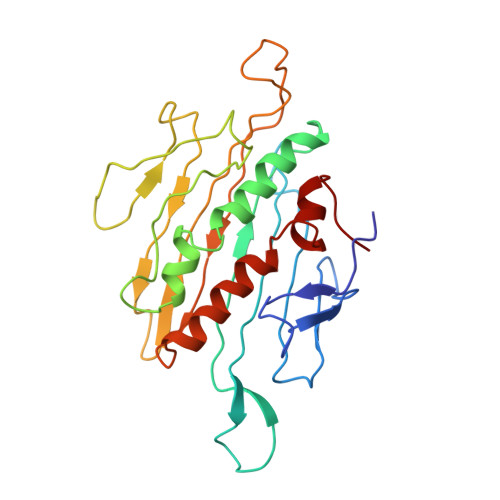
    A covalent dimer of the bacteriophage MS2 coat protein was created by performing genetic fusion of two copies of the gene while removing the stop codon of the first gene. The dimer was crystallized in the cubic F432 space group. The organization of the asymmetric unit together with the F432 symmetry results in an arrangement of subunits that corresponds to T = 3 octahedral particles. The octahedral particles are probably artifacts created by the particular crystal packing. When it is not crystallized in the F cubic crystal form, the coat protein dimer appears to assemble into T = 3 icosahedral particles indistinguishable from the wild-type particles. To form an octahedral particle with closed surface, the dimer subunits interact at sharper angles than in the icosahedral arrangement. The fold of the covalent dimer is almost identical to the wild-type dimer with differences located in loops and in the covalent linker region. The main differences in the subunit packing between the octahedral and icosahedral arrangements are located close to the fourfold and fivefold symmetry axes where different sets of loops mediate the contacts. The volume of the wild-type virions is 7 times bigger than that of the octahedral particles.


  • Organizational Affiliation

    Department of Cell and Molecular Biology, Uppsala University, SE-751 24 Uppsala, Sweden. pavel@xray.bmc.uu.se


Macromolecules
Find similar proteins by:  (by identity cutoff)  |  3D Structure
Entity ID: 1
MoleculeChains Sequence LengthOrganismDetailsImage
MS2 COAT PROTEINA [auth J],
B [auth L]
257Escherichia phage MS2Mutation(s): 0 
UniProt
Find proteins for P03612 (Escherichia phage MS2)
Explore P03612 
Go to UniProtKB:  P03612
Entity Groups  
Sequence Clusters30% Identity50% Identity70% Identity90% Identity95% Identity100% Identity
UniProt GroupP03612
Sequence Annotations
Expand
  • Reference Sequence
Experimental Data & Validation

Experimental Data

  • Method: X-RAY DIFFRACTION
  • Resolution: 3.50 Å
  • R-Value Free: 0.310 
  • R-Value Work: 0.272 
  • R-Value Observed: 0.272 
  • Space Group: F 4 3 2
Unit Cell:
Length ( Å )Angle ( ˚ )
a = 220.438α = 90
b = 220.438β = 90
c = 220.438γ = 90
Software Package:
Software NamePurpose
HKL-2000data reduction
SCALEPACKdata scaling
CNSphasing
BEASTphasing
CNSrefinement

Structure Validation

View Full Validation Report


Entry History 

Deposition Data

Revision History  (Full details and data files)

  • Version 1.0: 2008-08-12
    Type: Initial release
  • Version 1.1: 2011-07-07
    Changes: Version format compliance
  • Version 1.2: 2011-07-13
    Changes: Version format compliance
  • Version 1.3: 2023-12-13
    Changes: Data collection, Database references, Derived calculations, Other, Refinement description