2VQP

Structure of the matrix protein from human Respiratory Syncytial Virus


Experimental Data Snapshot

  • Method: X-RAY DIFFRACTION
  • Resolution: 1.60 Å
  • R-Value Free: 0.217 
  • R-Value Work: 0.180 
  • R-Value Observed: 0.182 

wwPDB Validation   3D Report Full Report


This is version 1.1 of the entry. See complete history


Literature

Surface Features of a Mononegavirales Matrix Protein Indicate Sites of Membrane Interaction.

Money, V.A.Mcphee, H.K.Mosely, J.A.Sanderson, J.M.Yeo, R.P.

(2009) Proc Natl Acad Sci U S A 106: 4441

  • DOI: https://doi.org/10.1073/pnas.0805740106
  • Primary Citation of Related Structures:  
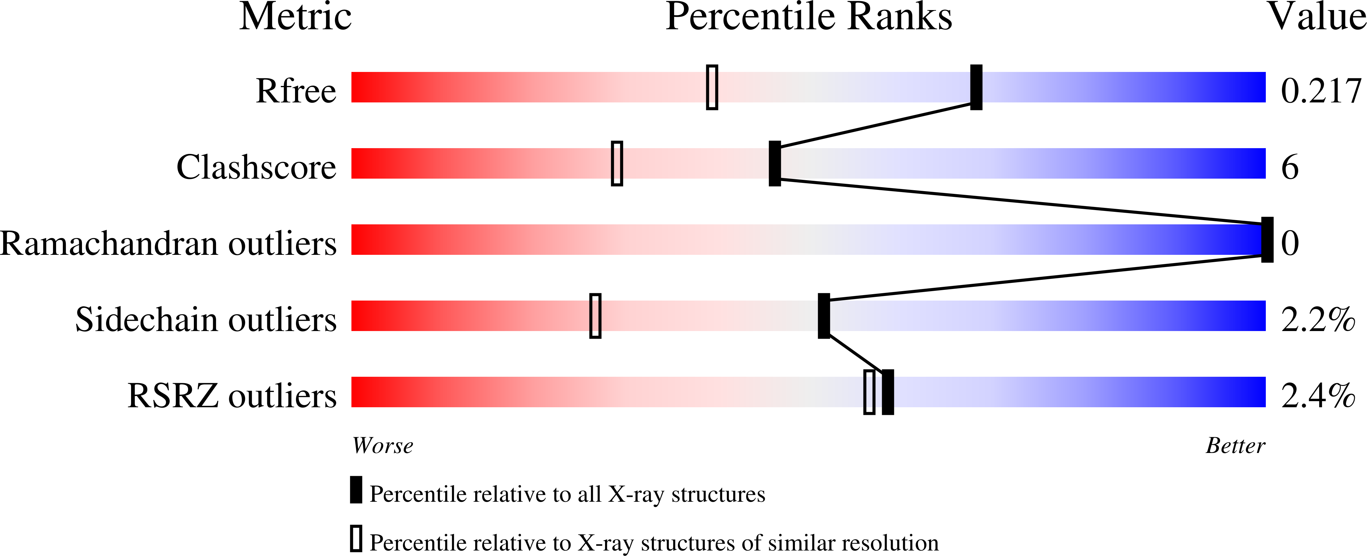
    2VQP

  • PubMed Abstract: 

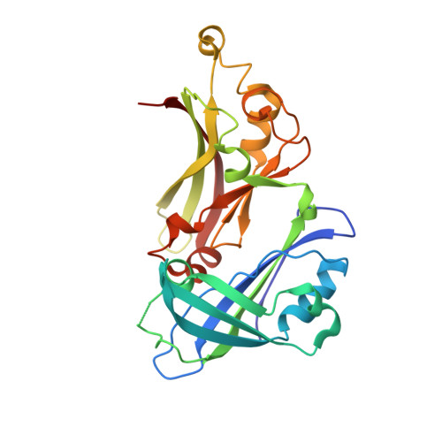
    The matrix protein (M) of respiratory syncytial virus (RSV), the prototype viral member of the Pneumovirinae (family Paramyxoviridae, order Mononegavirales), has been crystallized and the structure determined to a resolution of 1.6 A. The structure comprises 2 compact beta-rich domains connected by a relatively unstructured linker region. Due to the high degree of side-chain order in the structure, an extensive contiguous area of positive surface charge covering approximately 600 A(2) can be resolved. This unusually large patch of positive surface potential spans both domains and the linker, and provides a mechanism for driving the interaction of the protein with a negatively-charged membrane surface or other virion components such as the nucleocapsid. This patch is complemented by regions of high hydrophobicity and a striking planar arrangement of tyrosine residues encircling the C-terminal domain. Comparison of the RSV M sequence with other members of the Pneumovirinae shows that regions of divergence correspond to surface exposed loops in the M structure, with the majority of viral species-specific differences occurring in the N-terminal domain.


  • Organizational Affiliation

    The Maurice Wilkins Centre for Molecular Biodiscovery and the School of Biological Sciences, University of Auckland, Thomas Building, 3a Symonds Street, Auckland Central 1010, New Zealand.


Macromolecules
Find similar proteins by:  (by identity cutoff)  |  3D Structure
Entity ID: 1
MoleculeChains Sequence LengthOrganismDetailsImage
MATRIX PROTEIN257Human respiratory syncytial virus A2Mutation(s): 1 
Membrane Entity: Yes 
UniProt
Find proteins for P0DOE7 (Human respiratory syncytial virus A (strain A2))
Explore P0DOE7 
Go to UniProtKB:  P0DOE7
Entity Groups  
Sequence Clusters30% Identity50% Identity70% Identity90% Identity95% Identity100% Identity
UniProt GroupP0DOE7
Sequence Annotations
Expand
  • Reference Sequence
Small Molecules
Ligands 3 Unique
IDChains Name / Formula / InChI Key2D Diagram3D Interactions
GOL
Query on GOL

Download Ideal Coordinates CCD File 
B [auth A],
C [auth A]
GLYCEROL
C3 H8 O3
PEDCQBHIVMGVHV-UHFFFAOYSA-N
ACT
Query on ACT

Download Ideal Coordinates CCD File 
K [auth A],
L [auth A],
M [auth A]
ACETATE ION
C2 H3 O2
QTBSBXVTEAMEQO-UHFFFAOYSA-M
FMT
Query on FMT

Download Ideal Coordinates CCD File 
D [auth A]
E [auth A]
F [auth A]
G [auth A]
H [auth A]
D [auth A],
E [auth A],
F [auth A],
G [auth A],
H [auth A],
I [auth A],
J [auth A]
FORMIC ACID
C H2 O2
BDAGIHXWWSANSR-UHFFFAOYSA-N
Experimental Data & Validation

Experimental Data

  • Method: X-RAY DIFFRACTION
  • Resolution: 1.60 Å
  • R-Value Free: 0.217 
  • R-Value Work: 0.180 
  • R-Value Observed: 0.182 
  • Space Group: C 1 2 1
Unit Cell:
Length ( Å )Angle ( ˚ )
a = 52.299α = 90
b = 78.697β = 96.28
c = 66.018γ = 90
Software Package:
Software NamePurpose
REFMACrefinement
MOSFLMdata reduction
SCALAdata scaling
SHARPphasing

Structure Validation

View Full Validation Report


Entry History 

Deposition Data

Revision History  (Full details and data files)

  • Version 1.0: 2009-02-17
    Type: Initial release
  • Version 1.1: 2011-07-13
    Changes: Source and taxonomy, Version format compliance