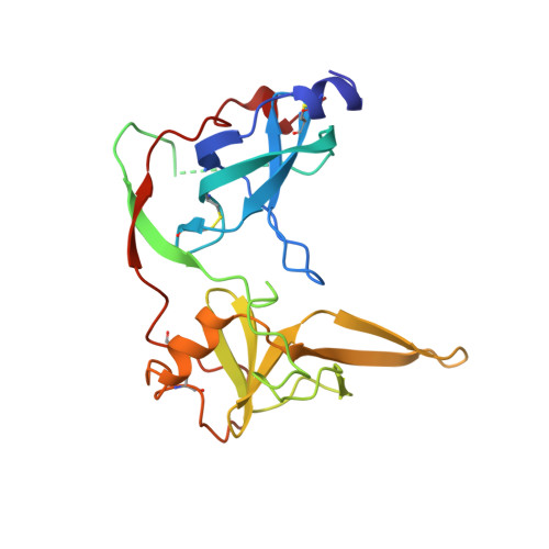
Crystal structure of the two N-terminal domains of g3p from filamentous phage fd at 1.9 A: evidence for conformational lability.
Holliger, P., Riechmann, L., Williams, R.L.(1999) J Mol Biol 288: 649-657
- PubMed: 10329170
- DOI: https://doi.org/10.1006/jmbi.1999.2720
- Primary Citation of Related Structures:
2G3P - PubMed Abstract:
Infection of Escherichia coli by filamentous bacteriophages is mediated by the minor phage coat protein g3p and involves two distinct cellular receptors, the F' pilus and the periplasmic protein TolA. Recently we have shown that the two receptors are contacted in a sequential manner, such that binding of TolA by the N-terminal domain g3p-D1 is conditional on a primary interaction of the second g3p domain D2 with the F' pilus. In order to better understand this process, we have solved the crystal structure of the g3p-D1D2 fragment (residues 2-217) from filamentous phage fd to 1.9 A resolution and compared it to the recently published structure of the same fragment from the related Ff phage M13. While the structure of individual domains D1 and D2 of the two phages are very similar (rms<0.7 A), there is comparatively poor agreement for the overall D1D2 structure (rms>1.2 A). This is due to an apparent movement of domain D2 with respect to D1, which results in a widening of the inter-domain groove compared to the structure of the homologous M13 protein. The movement of D2 can be described as a rigid-body rotation around a hinge located at the end of a short anti-parallel beta-sheet connecting domains D1 and D2. Structural flexibility of at least parts of the D1D2 structure was also suggested by studying the thermal unfolding of g3p: the TolA binding site on D1, while fully blocked by D2 at 37 degrees C, becomes accessible after incubation at temperatures as low as 45 degrees C. Our results support a model for the early steps of phage infection whereby exposure of the coreceptor binding site on D1 is facilitated by a conformational change in the D1D2 structure, which in vivo is induced by binding to the F' pilus on the host cell and which can be mimicked in vitro by thermal unfolding.
Organizational Affiliation:
MRC Centre for Protein Engineering, Cambridge CB2 2QH, UK. ph1@mrc-lmb.cam.ac.uk