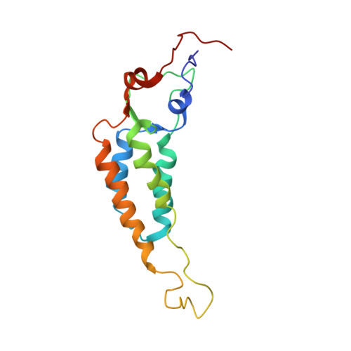
1VTM

STRUCTURE OF THE U2 STRAIN OF TOBACCO MOSAIC VIRUS REFINED AT 3.5 ANGSTROMS RESOLUTION USING X-RAY FIBER DIFFRACTION


Experimental Data Snapshot

  • Method: FIBER DIFFRACTION
  • Resolution: 3.50 Å
  • R-Value Observed: 0.096 

wwPDB Validation   3D Report Full Report


This is version 1.4 of the entry. See complete history


Literature

Structure of the U2 strain of tobacco mosaic virus refined at 3.5 A resolution using X-ray fiber diffraction.

Pattanayek, R.Stubbs, G.

(1992) J Mol Biol 228: 516-528

  • DOI: https://doi.org/10.1016/0022-2836(92)90839-c
  • Primary Citation of Related Structures:  
    1VTM

  • PubMed Abstract: 

    The structure of the U2 strain of tobacco mosaic virus (TMV) has been determined by fiber diffraction methods at 3.5 A resolution, and refined by a combination of restrained least-squares and molecular dynamics methods to an R-factor of 0.096. The structure is extremely similar to that of the common strain of TMV, with the largest differences being in the protein loop that makes up the inner surface of the virus, and in the C-terminal region on the outer surface. Differences in the inner loop can be correlated with differences in the properties of the two viruses.


  • Organizational Affiliation

    Department of Molecular Biology Vanderbilt University, Nashville, TN 37235.


Macromolecules

Find similar proteins by:  (by identity cutoff)  |  3D Structure
Entity ID: 2
MoleculeChains Sequence LengthOrganismDetailsImage
Coat proteinB [auth P]158Tobacco mild green mosaic virusMutation(s): 0 
Gene Names: CP
UniProt
Find proteins for P03579 (Tobacco mild green mosaic virus)
Explore P03579 
Go to UniProtKB:  P03579
Entity Groups  
Sequence Clusters30% Identity50% Identity70% Identity90% Identity95% Identity100% Identity
UniProt GroupP03579
Sequence Annotations
Expand
  • Reference Sequence

Find similar nucleic acids by:  Sequence   |   3D Structure  

Entity ID: 1
MoleculeChains LengthOrganismImage
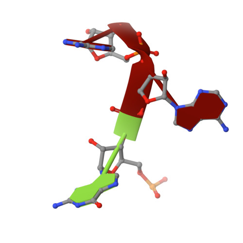
RNA (5'-R(P*GP*AP*A)-3')A [auth R]3Tobacco mild green mosaic virus
Sequence Annotations
Expand
  • Reference Sequence
Experimental Data & Validation

Experimental Data

  • Method: FIBER DIFFRACTION
  • Resolution: 3.50 Å
  • R-Value Observed: 0.096 
  • Space Group: P 1
Unit Cell:
Length ( Å )Angle ( ˚ )
a = 1α = 90
b = 1β = 90
c = 1γ = 90
Software Package:
Software NamePurpose
X-PLORmodel building
PROLSQrefinement
X-PLORrefinement
X-PLORphasing

Structure Validation

View Full Validation Report


Entry History 

Deposition Data

Revision History  (Full details and data files)

  • Version 1.0: 1994-07-31
    Type: Initial release
  • Version 1.1: 2008-03-03
    Changes: Version format compliance
  • Version 1.2: 2011-07-13
    Changes: Version format compliance
  • Version 1.3: 2017-11-29
    Changes: Derived calculations, Other
  • Version 1.4: 2024-02-14
    Changes: Data collection, Database references, Derived calculations