


X-ray crystallographic studies of archaerhodopsin
Enami, N., Okumua, H., Kouyama, T.(2003) J Photosci 9: 320-322
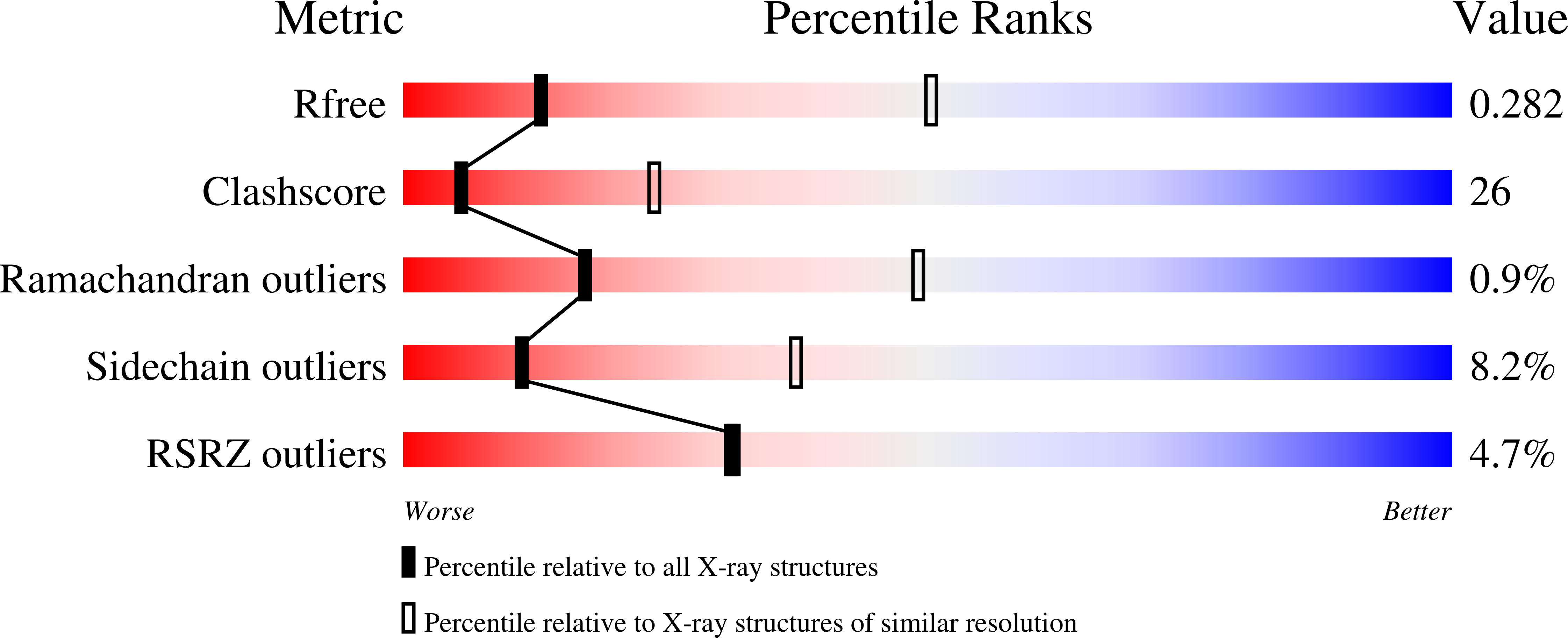
Experimental Data Snapshot
(2003) J Photosci 9: 320-322
Entity ID: 1 | |||||
|---|---|---|---|---|---|
| Molecule | Chains | Sequence Length | Organism | Details | Image |
| archaerhodopsin-1 | 254 | Halorubrum ezzemoulense | Mutation(s): 0 Membrane Entity: Yes | 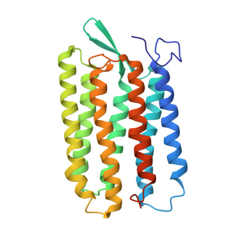 | |
UniProt | |||||
Find proteins for P69051 (Halorubrum ezzemoulense) Explore P69051 Go to UniProtKB: P69051 | |||||
Entity Groups | |||||
| Sequence Clusters | 30% Identity50% Identity70% Identity90% Identity95% Identity100% Identity | ||||
| UniProt Group | P69051 | ||||
Sequence AnnotationsExpand | |||||
| |||||
| Ligands 1 Unique | |||||
|---|---|---|---|---|---|
| ID | Chains | Name / Formula / InChI Key | 2D Diagram | 3D Interactions | |
| RET Query on RET | C [auth A], D [auth B] | RETINAL C20 H28 O NCYCYZXNIZJOKI-OVSJKPMPSA-N |  | ||
| Length ( Å ) | Angle ( ˚ ) |
|---|---|
| a = 128.1 | α = 90 |
| b = 128.1 | β = 90 |
| c = 117.6 | γ = 90 |
| Software Name | Purpose |
|---|---|
| MOSFLM | data reduction |
| SCALA | data scaling |
| XTALVIEW | refinement |
| CNS | refinement |
| CCP4 | data scaling |
RCSB PDB (citation) is hosted by
RCSB PDB is a member of the

