Structure and Biochemical Features Distinguish the Foot-and-Mouth Disease Virus Leader Proteinase from Other Papain-Like Enzymes
Guarne, A., Hampoelz, B., Glaser, W., Carpena, X., Tormo, J., Fita, I., Skern, T.(2000) J Mol Biol 302: 1227
- PubMed: 11183785
- DOI: https://doi.org/10.1006/jmbi.2000.4115
- Primary Citation of Related Structures:
1QMY - PubMed Abstract:
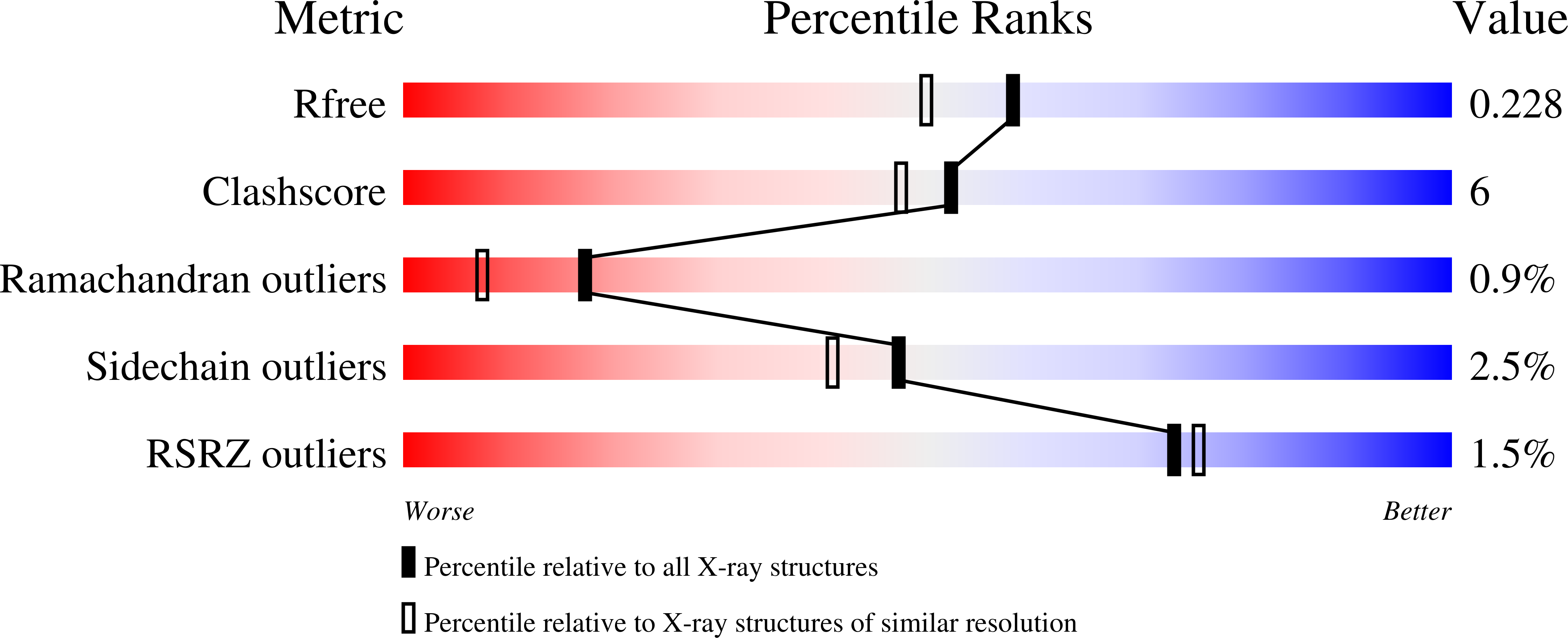
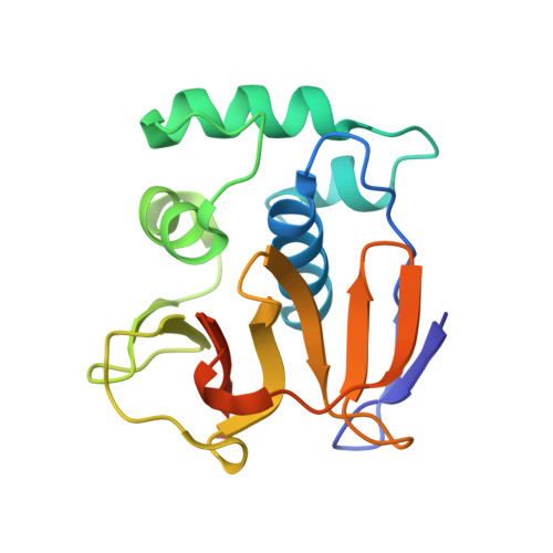
The structures of the two leader protease (Lpro) variants of foot-and-mouth disease virus known to date were solved using crystals in which molecules were organized as molecular fibers. Such crystals diffract to a resolution of only approximately 3 A. This singular, pseudo-polymeric organization is present in a new Lpro crystal form showing a cubic packing. As molecular fiber formation appeared unrelated to crystallization conditions, we mutated the reactive cysteine 133 residue, which makes a disulfide bridge between adjacent monomers in the fibers, to serine. None of the intermolecular contacts found in the molecular fibers was present in crystals of this variant. Analysis of this Lpro structure, refined at 1.9 A resolution, enables a detailed definition of the active center of the enzyme, including the solvent organization. Assay of Lpro activity on a fluorescent hexapeptide substrate showed that Lpro, in contrast to papain, was highly sensitive to increases in the cation concentration and was active only across a narrow pH range. Examination of the Lpro structure revealed that three aspartate residues near the active site, not present in papain-like enzymes, are probably responsible for these properties.
Organizational Affiliation:
Institut de Biologia Molecular de Barcelona, Barcelona, Spain.


