Structure-based design and in-parallel synthesis of inhibitors of AmpC beta-lactamase.
Tondi, D., Powers, R.A., Caselli, E., Negri, M.C., Blazquez, J., Costi, M.P., Shoichet, B.K.(2001) Chem Biol 8: 593-611
- PubMed: 11410378
- DOI: https://doi.org/10.1016/s1074-5521(01)00034-5
- Primary Citation of Related Structures:
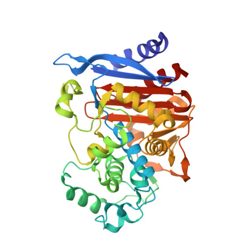
1GA9 - PubMed Abstract:
Group I beta-lactamases are a major cause of antibiotic resistance to beta-lactams such as penicillins and cephalosporins. These enzymes are only modestly affected by classic beta-lactam-based inhibitors, such as clavulanic acid. Conversely, small arylboronic acids inhibit these enzymes at sub-micromolar concentrations. Structural studies suggest these inhibitors bind to a well-defined cleft in the group I beta-lactamase AmpC; this cleft binds the ubiquitous R1 side chain of beta-lactams. Intriguingly, much of this cleft is left unoccupied by the small arylboronic acids. To investigate if larger boronic acids might take advantage of this cleft, structure-guided in-parallel synthesis was used to explore new inhibitors of AmpC. Twenty-eight derivatives of the lead compound, 3-aminophenylboronic acid, led to an inhibitor with 80-fold better binding (2; K(i) 83 nM). Molecular docking suggested orientations for this compound in the R1 cleft. Based on the docking results, 12 derivatives of 2 were synthesized, leading to inhibitors with K(i) values of 60 nM and with improved solubility. Several of these inhibitors reversed the resistance of nosocomial Gram-positive bacteria, though they showed little activity against Gram-negative bacteria. The X-ray crystal structure of compound 2 in complex with AmpC was subsequently determined to 2.1 A resolution. The placement of the proximal two-thirds of the inhibitor in the experimental structure corresponds with the docked structure, but a bond rotation leads to a distinctly different placement of the distal part of the inhibitor. In the experimental structure, the inhibitor interacts with conserved residues in the R1 cleft whose role in recognition has not been previously explored. Combining structure-based design with in-parallel synthesis allowed for the rapid exploration of inhibitor functionality in the R1 cleft of AmpC. The resulting inhibitors differ considerably from beta-lactams but nevertheless inhibit the enzyme well. The crystal structure of 2 (K(i) 83 nM) in complex with AmpC may guide exploration of a highly conserved, largely unexplored cleft, providing a template for further design against AmpC beta-lactamase.
Organizational Affiliation:
Department of Molecular Pharmacology and Biological Chemistry, Northwestern University, Chicago, IL 60611, USA.