Crystal structures of Bacillus caldovelox arginase in complex with substrate and inhibitors reveal new insights into activation, inhibition and catalysis in the arginase superfamily.
Bewley, M.C., Jeffrey, P.D., Patchett, M.L., Kanyo, Z.F., Baker, E.N.(1999) Structure 7: 435-448
- PubMed: 10196128
- DOI: https://doi.org/10.1016/s0969-2126(99)80056-2
- Primary Citation of Related Structures:
1CEV, 2CEV, 3CEV, 4CEV, 5CEV - PubMed Abstract:
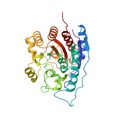
Arginase is a manganese-dependent enzyme that catalyzes the hydrolysis of L-arginine to L-ornithine and urea. In ureotelic animals arginase is the final enzyme of the urea cycle, but in many species it has a wider role controlling the use of arginine for other metabolic purposes, including the production of creatine, polyamines, proline and nitric oxide. Arginase activity is regulated by various small molecules, including the product L-ornithine. The aim of these structural studies was to test aspects of the catalytic mechanism and to investigate the structural basis of arginase inhibition. We report here the crystal structures of arginase from Bacillus caldovelox at pH 5.6 and pH 8.5, and of binary complexes of the enzyme with L-arginine, L-ornithine and L-lysine at pH 8.5. The arginase monomer comprises a single compact alpha/beta domain that further associates into a hexameric quaternary structure. The binary complexes reveal a common mode of ligand binding, which places the substrate adjacent to the dimanganese centre. We also observe a conformational change that impacts on the active site and is coupled with the occupancy of an external site by guanidine or arginine. The structures reported here clarify aspects of the active site and indicate key features of the catalytic mechanism, including substrate coordination to one of the manganese ions and an orientational role for a neighboring histidine residue. Stereospecificity for L-amino acids is found to depend on their precise recognition at the active-site rim. Identification of a second arginine-binding site, remote from the active site, and associated conformational changes lead us to propose a regulatory role for this site in substrate hydrolysis.
Organizational Affiliation:
Department of Biochemistry, Massey University, Palmerston North, New Zealand. bewley@js1.bio.bnl.gov.