Protein engineering of xylose (glucose) isomerase from Actinoplanes missouriensis. 1. Crystallography and site-directed mutagenesis of metal binding sites.
Jenkins, J., Janin, J., Rey, F., Chiadmi, M., van Tilbeurgh, H., Lasters, I., De Maeyer, M., Van Belle, D., Wodak, S.J., Lauwereys, M., Stanssens, P., Matthyssens, G., Lambeir, A.M.(1992) Biochemistry 31: 5449-5458
- PubMed: 1610791
- DOI: https://doi.org/10.1021/bi00139a005
- Primary Citation of Related Structures:
1XIN, 2XIN, 3XIN, 4XIM, 5XIM, 5XIN, 6XIM, 7XIM, 8XIM, 9XIM - PubMed Abstract:
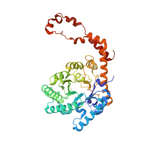
The structure and function of the xylose (glucose) isomerase from Actinoplanes missouriensis have been analyzed by X-ray crystallography and site-directed mutagenesis after cloning and overexpression in Escherichia coli. The crystal structure of wild-type enzyme has been refined to an R factor of 15.2% against diffraction data to 2.2-A resolution. The structures of a number of binary and ternary complexes involving wild-type and mutant enzymes, the divalent cations Mg2+, Co2+, or Mn2+, and either the substrate xylose or substrate analogs have also been determined and refined to comparable R factors. Two metal sites are identified. Metal site 1 is four-coordinated and tetrahedral in the absence of substrate and is six-coordinated and octahedral in its presence; the O2 and O4 atoms of linear inhibitors and substrate bind to metal 1. Metal site 2 is octahedral in all cases; its position changes by 0.7 A when it binds O1 of the substrate and by more than 1 A when it also binds O2; these bonds replace bonds to carboxylate ligands from the protein. Side chains involved in metal binding have been substituted by site-directed mutagenesis. The biochemical properties of the mutant enzymes are presented. Together with structural data, they demonstrate that the two metal ions play an essential part in binding substrates, in stabilizing their open form, and in catalyzing hydride transfer between the C1 and C2 positions.
Organizational Affiliation:
Laboratoire de Biologie Physicochimique, CNRS UA1131, Université Paris-Sud, Orsay, France.