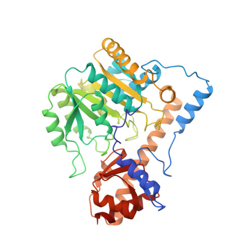
X-ray structure refinement and comparison of three forms of mitochondrial aspartate aminotransferase.
McPhalen, C.A., Vincent, M.G., Jansonius, J.N.(1992) J Mol Biol 225: 495-517
- PubMed: 1593633
- DOI: https://doi.org/10.1016/0022-2836(92)90935-d
- Primary Citation of Related Structures:
7AAT, 8AAT, 9AAT - PubMed Abstract:
The X-ray crystal structures of three forms of the enzyme aspartate aminotransferase (EC 2.6.1.1) from chicken heart mitochondria have been refined by least-squares methods: holoenzyme with the co-factor pyridoxal-5'-phosphate bound at pH 7.5 (1.9 A resolution), holoenzyme with pyridoxal-5'-phosphate bound at pH 5.1 (2.3 A resolution) and holoenzyme with the co-factor pyridoxamine-5'-phosphate bound at pH 7.5 (2.2 A resolution). The crystallographic agreement factors [formula: see text] for the structures are 0.166, 0.130 and 0.131, respectively, for all data in the resolution range from 10.0 A to the limit of diffraction for each structure. The secondary, super-secondary and domain structures of the pyridoxal-phosphate holoenzyme at pH 7.5 are described in detail. The surface area of the interface between the monomer subunits of this dimeric alpha 2 protein is unusually large, indicating a very stable dimer. This is consistent with biochemical data. Both subunit and domain interfaces are relatively smooth compared with other proteins. The interactions of the protein with its co-factor are described and compared among the three structures. Observed changes in co-factor conformation may be related to spectral changes and the energetics of the catalytic reaction. Small but significant adjustments of the protein to changes in co-factor conformation are seen. These adjustments may be accommodated by small rigid-body shifts of secondary structural elements, and by packing defects in the protein core.
Organizational Affiliation:
Department of Structural Biology, University of Basel, Switzerland.


