Caenorhabditis elegans telomere-binding proteins TEBP-1 and TEBP-2 adapt the Myb module to dimerize and bind telomeric DNA.
Padmanaban, S., Lambacher, N.J., Tesmer, V.M., Zhang, J., Shibuya, H., Nandakumar, J.(2024) Proc Natl Acad Sci U S A 121: e2316651121-e2316651121
- PubMed: 38588418
- DOI: https://doi.org/10.1073/pnas.2316651121
- Primary Citation of Related Structures:
8U8L, 8U8M - PubMed Abstract:
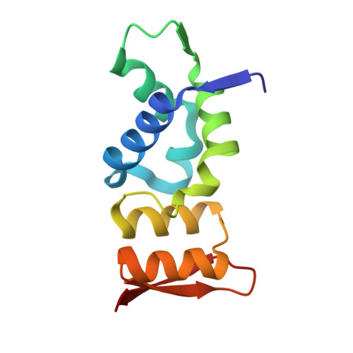
Protecting chromosome ends from misrecognition as double-stranded (ds) DNA breaks is fundamental to eukaryotic viability. The protein complex shelterin prevents a DNA damage response at mammalian telomeres. Mammalian shelterin proteins TRF1 and TRF2 and their homologs in yeast and protozoa protect telomeric dsDNA. N-terminal homodimerization and C-terminal Myb-domain-mediated dsDNA binding are two structural hallmarks of end protection by TRF homologs. Yet our understanding of how Caenorhabditis elegans protects its telomeric dsDNA is limited. Recently identified C. elegans proteins TEBP-1 (also called DTN-1) and TEBP-2 (also called DTN-2) are functional homologs of TRF proteins, but how they bind DNA and whether or how they dimerize is not known. TEBP-1 and TEBP-2 harbor three Myb-containing domains (MCDs) and no obvious dimerization domain. We demonstrate biochemically that only the third MCD binds DNA. We solve the X-ray crystal structure of TEBP-2 MCD3 with telomeric dsDNA to reveal the structural mechanism of telomeric dsDNA protection in C. elegans . Mutagenesis of the DNA-binding site of TEBP-1 and TEBP-2 compromises DNA binding in vitro, and increases DNA damage signaling, lengthens telomeres, and decreases brood size in vivo. Via an X-ray crystal structure, biochemical validation of the dimerization interface, and SEC-MALS analysis, we demonstrate that MCD1 and MCD2 form a composite dimerization module that facilitates not only TEBP-1 and TEBP-2 homodimerization but also heterodimerization. These findings provide fundamental insights into C. elegans telomeric dsDNA protection and highlight how different eukaryotes have evolved distinct strategies to solve the chromosome end protection problem.
Organizational Affiliation:
Department of Molecular, Cellular, and Developmental Biology, University of Michigan, Ann Arbor, MI 48109.