8TWD

Structure of IraM-bound RssB


Experimental Data Snapshot

  • Method: X-RAY DIFFRACTION
  • Resolution: 3.30 Å
  • R-Value Free: 0.271 
  • R-Value Work: 0.245 
  • R-Value Observed: 0.247 

wwPDB Validation   3D Report Full Report


This is version 1.1 of the entry. See complete history


Literature

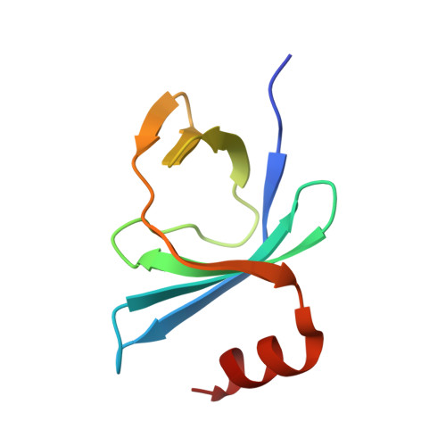
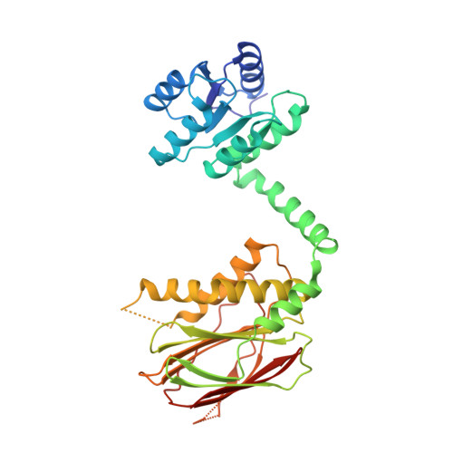
IraM remodels the RssB segmented helical linker to stabilize sigma s against degradation by ClpXP.

Brugger, C.Srirangam, S.Deaconescu, A.M.

(2023) J Biol Chem 300: 105568-105568

  • DOI: https://doi.org/10.1016/j.jbc.2023.105568
  • Primary Citation of Related Structures:  
    8TWD

  • PubMed Abstract: 

    Upon Mg 2+ starvation, a condition often associated with virulence, enterobacteria inhibit the ClpXP-dependent proteolysis of the master transcriptional regulator, σ s , via IraM, a poorly understood antiadaptor that prevents RssB-dependent loading of σ s onto ClpXP. This inhibition results in σ s accumulation and expression of stress resistance genes. Here, we report on the structural analysis of RssB bound to IraM, which reveals that IraM induces two folding transitions within RssB, amplified via a segmented helical linker. These conformational changes result in an open, yet inhibited RssB structure in which IraM associates with both the C-terminal and N-terminal domains of RssB and prevents binding of σ s to the 4-5-5 face of the N-terminal receiver domain. This work highlights the remarkable structural plasticity of RssB and reveals how a stress-specific RssB antagonist modulates a core stress response pathway that could be leveraged to control biofilm formation, virulence, and the development of antibiotic resistance.


  • Organizational Affiliation

    Department of Molecular Biology, Cell Biology and Biochemistry, Brown University, Providence, Rhode Island, USA.


Macromolecules
Find similar proteins by:  (by identity cutoff)  |  3D Structure
Entity ID: 1
MoleculeChains Sequence LengthOrganismDetailsImage
Anti-adapter protein IraM
A, B
81Escherichia coliMutation(s): 0 
Gene Names: iraM
UniProt
Find proteins for P75987 (Escherichia coli (strain K12))
Explore P75987 
Go to UniProtKB:  P75987
Entity Groups  
Sequence Clusters30% Identity50% Identity70% Identity90% Identity95% Identity100% Identity
UniProt GroupP75987
Sequence Annotations
Expand
  • Reference Sequence
Find similar proteins by:  (by identity cutoff)  |  3D Structure
Entity ID: 2
MoleculeChains Sequence LengthOrganismDetailsImage
Regulator of RpoS
C, D
337Escherichia coliMutation(s): 0 
Gene Names: rssBhnrsprEychLb1235JW1223
UniProt
Find proteins for P0AEV1 (Escherichia coli (strain K12))
Explore P0AEV1 
Go to UniProtKB:  P0AEV1
Entity Groups  
Sequence Clusters30% Identity50% Identity70% Identity90% Identity95% Identity100% Identity
UniProt GroupP0AEV1
Sequence Annotations
Expand
  • Reference Sequence
Small Molecules
Ligands 1 Unique
IDChains Name / Formula / InChI Key2D Diagram3D Interactions
ACT
Query on ACT

Download Ideal Coordinates CCD File 
E [auth A]ACETATE ION
C2 H3 O2
QTBSBXVTEAMEQO-UHFFFAOYSA-M
Experimental Data & Validation

Experimental Data

  • Method: X-RAY DIFFRACTION
  • Resolution: 3.30 Å
  • R-Value Free: 0.271 
  • R-Value Work: 0.245 
  • R-Value Observed: 0.247 
  • Space Group: C 2 2 21
Unit Cell:
Length ( Å )Angle ( ˚ )
a = 130.664α = 90
b = 191.229β = 90
c = 96.985γ = 90
Software Package:
Software NamePurpose
PHENIXrefinement
XDSdata reduction
Aimlessdata scaling
PHASERphasing

Structure Validation

View Full Validation Report


Entry History & Funding Information

Deposition Data


Funding OrganizationLocationGrant Number
National Institutes of Health/National Institute of General Medical Sciences (NIH/NIGMS)United StatesR01GM121975
National Institutes of Health/National Institute of General Medical Sciences (NIH/NIGMS)United StatesR35GM144124

Revision History  (Full details and data files)

  • Version 1.0: 2024-01-10
    Type: Initial release
  • Version 1.1: 2024-02-14
    Changes: Database references