8TKP

Structure of the C. elegans TMC-2 complex


Experimental Data Snapshot

  • Method: ELECTRON MICROSCOPY
  • Resolution: 2.90 Å
  • Aggregation State: TISSUE 
  • Reconstruction Method: SINGLE PARTICLE 

wwPDB Validation   3D Report Full Report


This is version 1.0 of the entry. See complete history


Literature

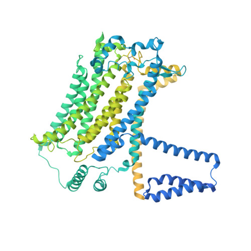
The structure of the Caenorhabditis elegans TMC-2 complex suggests roles of lipid-mediated subunit contacts in mechanosensory transduction.

Clark, S.Jeong, H.Posert, R.Goehring, A.Gouaux, E.

(2024) Proc Natl Acad Sci U S A 121: e2314096121-e2314096121

  • DOI: https://doi.org/10.1073/pnas.2314096121
  • Primary Citation of Related Structures:  
    8TKP

  • PubMed Abstract: 

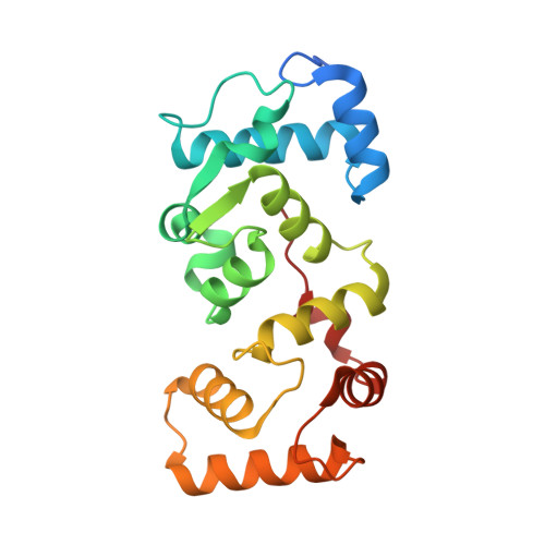
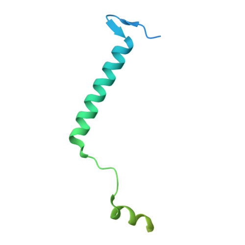
    Mechanotransduction is the process by which a mechanical force, such as touch, is converted into an electrical signal. Transmembrane channel-like (TMC) proteins are an evolutionarily conserved family of membrane proteins whose function has been linked to a variety of mechanosensory processes, including hearing and balance sensation in vertebrates and locomotion in Drosophila . TMC1 and TMC2 are components of ion channel complexes, but the molecular features that tune these complexes to diverse mechanical stimuli are unknown. Caenorhabditis elegans express two TMC homologs, TMC-1 and TMC-2, both of which are the likely pore-forming subunits of mechanosensitive ion channels but differ in their expression pattern and functional role in the worm. Here, we present the single-particle cryo-electron microscopy structure of the native TMC-2 complex isolated from C. elegans . The complex is composed of two copies of the pore-forming TMC-2 subunit, the calcium and integrin binding protein CALM-1 and the transmembrane inner ear protein TMIE. Comparison of the TMC-2 complex to the recently published cryo-EM structure of the C. elegans TMC-1 complex highlights conserved protein-lipid interactions, as well as a π-helical structural motif in the pore-forming helices, that together suggest a mechanism for TMC-mediated mechanosensory transduction.


  • Organizational Affiliation

    Vollum Institute, Oregon Health and Science University, Portland, OR 97239.


Macromolecules
Find similar proteins by:  (by identity cutoff)  |  3D Structure
Entity ID: 1
MoleculeChains Sequence LengthOrganismDetailsImage
Transmembrane channel-like protein 2
A, D
1,203Caenorhabditis elegansMutation(s): 0 
Gene Names: tmc-2B0416.1
Membrane Entity: Yes 
UniProt
Find proteins for Q11069 (Caenorhabditis elegans)
Explore Q11069 
Go to UniProtKB:  Q11069
Entity Groups  
Sequence Clusters30% Identity50% Identity70% Identity90% Identity95% Identity100% Identity
UniProt GroupQ11069
Sequence Annotations
Expand
  • Reference Sequence
Find similar proteins by:  (by identity cutoff)  |  3D Structure
Entity ID: 2
MoleculeChains Sequence LengthOrganismDetailsImage
CALMyrin (Calcium and Integrin Binding protein) homolog
B, E
201Caenorhabditis elegansMutation(s): 0 
Gene Names: calm-1CELE_F30A10.1F30A10.1
UniProt
Find proteins for Q93640 (Caenorhabditis elegans)
Explore Q93640 
Go to UniProtKB:  Q93640
Entity Groups  
Sequence Clusters30% Identity50% Identity70% Identity90% Identity95% Identity100% Identity
UniProt GroupQ93640
Sequence Annotations
Expand
  • Reference Sequence
Find similar proteins by:  (by identity cutoff)  |  3D Structure
Entity ID: 3
MoleculeChains Sequence LengthOrganismDetailsImage
Transmembrane inner ear expressed protein
C, F
117Caenorhabditis elegansMutation(s): 0 
Gene Names: CELE_Y39A1C.1Y39A1C.1
Membrane Entity: Yes 
UniProt
Find proteins for Q9XXE7 (Caenorhabditis elegans)
Explore Q9XXE7 
Go to UniProtKB:  Q9XXE7
Entity Groups  
Sequence Clusters30% Identity50% Identity70% Identity90% Identity95% Identity100% Identity
UniProt GroupQ9XXE7
Sequence Annotations
Expand
  • Reference Sequence
Oligosaccharides

Help

Entity ID: 4
MoleculeChains Length2D Diagram Glycosylation3D Interactions
2-acetamido-2-deoxy-beta-D-glucopyranose-(1-3)-2-acetamido-2-deoxy-beta-D-glucopyranoseG [auth H]2N-Glycosylation
Glycosylation Resources
GlyTouCan:  G79924RM
GlyCosmos:  G79924RM
GlyGen:  G79924RM
Entity ID: 5
MoleculeChains Length2D Diagram Glycosylation3D Interactions
2-acetamido-2-deoxy-beta-D-glucopyranose-(1-4)-2-acetamido-2-deoxy-beta-D-glucopyranoseH [auth G]2N-Glycosylation
Glycosylation Resources
GlyTouCan:  G42666HT
GlyCosmos:  G42666HT
GlyGen:  G42666HT
Small Molecules
Ligands 7 Unique
IDChains Name / Formula / InChI Key2D Diagram3D Interactions
PEE
Query on PEE

Download Ideal Coordinates CCD File 
FA [auth C]
IA [auth D]
J [auth A]
RA [auth D]
S [auth A]
FA [auth C],
IA [auth D],
J [auth A],
RA [auth D],
S [auth A],
SA [auth D],
T [auth A],
UA [auth D],
V [auth A],
XA [auth D]
1,2-dioleoyl-sn-glycero-3-phosphoethanolamine
C41 H78 N O8 P
MWRBNPKJOOWZPW-NYVOMTAGSA-N
CLR
Query on CLR

Download Ideal Coordinates CCD File 
LA [auth D],
M [auth A]
CHOLESTEROL
C27 H46 O
HVYWMOMLDIMFJA-DPAQBDIFSA-N
TWT
Query on TWT

Download Ideal Coordinates CCD File 
BA [auth C]
BB [auth F]
HA [auth D]
I [auth A]
VA [auth D]
BA [auth C],
BB [auth F],
HA [auth D],
I [auth A],
VA [auth D],
W [auth A]
DOCOSANE
C22 H46
HOWGUJZVBDQJKV-UHFFFAOYSA-N
PLM (Subject of Investigation/LOI)
Query on PLM

Download Ideal Coordinates CCD File 
EA [auth C],
EB [auth F],
FB [auth F],
GA [auth C]
PALMITIC ACID
C16 H32 O2
IPCSVZSSVZVIGE-UHFFFAOYSA-N
R16
Query on R16

Download Ideal Coordinates CCD File 
CA [auth C]
CB [auth F]
DA [auth C]
DB [auth F]
JA [auth D]
CA [auth C],
CB [auth F],
DA [auth C],
DB [auth F],
JA [auth D],
K [auth A],
KA [auth D],
L [auth A],
MA [auth D],
N [auth A],
NA [auth D],
O [auth A],
OA [auth D],
P [auth A],
PA [auth D],
Q [auth A],
QA [auth D],
R [auth A],
TA [auth D],
U [auth A],
Y [auth A],
YA [auth D]
HEXADECANE
C16 H34
DCAYPVUWAIABOU-UHFFFAOYSA-N
NAG
Query on NAG

Download Ideal Coordinates CCD File 
WA [auth D],
X [auth A]
2-acetamido-2-deoxy-beta-D-glucopyranose
C8 H15 N O6
OVRNDRQMDRJTHS-FMDGEEDCSA-N
CA
Query on CA

Download Ideal Coordinates CCD File 
AA [auth B],
AB [auth E],
Z [auth B],
ZA [auth E]
CALCIUM ION
Ca
BHPQYMZQTOCNFJ-UHFFFAOYSA-N
Experimental Data & Validation

Experimental Data

  • Method: ELECTRON MICROSCOPY
  • Resolution: 2.90 Å
  • Aggregation State: TISSUE 
  • Reconstruction Method: SINGLE PARTICLE 

Structure Validation

View Full Validation Report


Entry History & Funding Information

Deposition Data


Funding OrganizationLocationGrant Number
Howard Hughes Medical Institute (HHMI)United States--

Revision History  (Full details and data files)

  • Version 1.0: 2024-03-27
    Type: Initial release