8T7J

Oxygen- and PLP-dependent Cap15 holoenzyme bound with phosphate anion


Experimental Data Snapshot

  • Method: X-RAY DIFFRACTION
  • Resolution: 2.40 Å
  • R-Value Free: 0.268 
  • R-Value Work: 0.224 
  • R-Value Observed: 0.226 

wwPDB Validation   3D Report Full Report


Ligand Structure Quality Assessment 


This is version 1.2 of the entry. See complete history


Literature

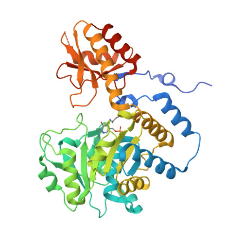
Structure of the Oxygen, Pyridoxal Phosphate-Dependent Capuramycin Biosynthetic Protein Cap15.

Daniel-Ivad, P.G.Van Lanen, S.Ryan, K.S.

(2023) Biochemistry 62: 2611-2621

  • DOI: https://doi.org/10.1021/acs.biochem.3c00216
  • Primary Citation of Related Structures:  
    8T7J

  • PubMed Abstract: 

    Pyridoxal phosphate-dependent enzymes able to use oxygen as a co-substrate have emerged in multiple protein families. Here, we use crystallography to solve the 2.40 Å resolution crystal structure of Cap15, a nucleoside biosynthetic enzyme that catalyzes the oxidative decarboxylation of glycyl uridine. Our structural study captures the internal aldimine, pinpointing the active site lysine as K230 and showing the site of phosphate binding. Our docking studies reveal how Cap15 is able to catalyze a stereoselective deprotonation reaction, and bioinformatic analysis reveals active site residues that distinguish Cap15 from the structurally related d-glucosaminate-6-phosphate ammonia lyase and l-seryl-tRNA(Sec) selenium transferase (SelA). Our work provides the structural basis for further mechanistic investigation of a unique biosynthetic enzyme and provides a blueprint for understanding how oxygen reactivity emerged in the SelA-like protein family.


  • Organizational Affiliation

    Department of Chemistry, University of British Columbia, Vancouver, British Columbia V6T 1Z1, Canada.


Macromolecules
Find similar proteins by:  (by identity cutoff)  |  3D Structure
Entity ID: 1
MoleculeChains Sequence LengthOrganismDetailsImage
Putative selenocysteine synthase
A, B, C, D
399Streptomyces sp. SANK 62799Mutation(s): 0 
UniProt
Find proteins for E1CG36 (Streptomyces sp)
Explore E1CG36 
Go to UniProtKB:  E1CG36
Entity Groups  
Sequence Clusters30% Identity50% Identity70% Identity90% Identity95% Identity100% Identity
UniProt GroupE1CG36
Sequence Annotations
Expand
  • Reference Sequence
Experimental Data & Validation

Experimental Data

  • Method: X-RAY DIFFRACTION
  • Resolution: 2.40 Å
  • R-Value Free: 0.268 
  • R-Value Work: 0.224 
  • R-Value Observed: 0.226 
  • Space Group: C 2 2 21
Unit Cell:
Length ( Å )Angle ( ˚ )
a = 142.59α = 90
b = 220.986β = 90
c = 96.735γ = 90
Software Package:
Software NamePurpose
PHENIXrefinement
Blu-Icedata collection
XDSdata reduction
Aimlessdata scaling
PHASERphasing
Cootmodel building

Structure Validation

View Full Validation Report


Ligand Structure Quality Assessment 


Entry History & Funding Information

Deposition Data


Funding OrganizationLocationGrant Number
Not funded--

Revision History  (Full details and data files)

  • Version 1.0: 2023-08-23
    Type: Initial release
  • Version 1.1: 2023-09-13
    Changes: Database references
  • Version 1.2: 2023-11-15
    Changes: Data collection